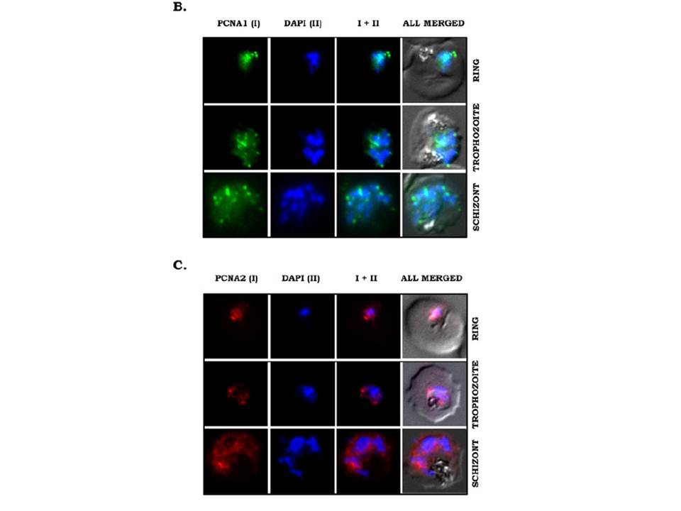
Stage-specific expression and subcellular localization of PfPCNA proteins. (B and C) IFA was performed on glass slides containing parasite smears, which were treated with antibodies against PfPCNA1 (B) or PfPCNA2 (C). PfPCNA1 (green) shows nuclear foci formation in replicating trophozoite stage and multinucleate schizont stage, whereas PfPCNA2 (red) shows both nuclear and cytoplasmic diffuse staining. DAPI (blue) stains the nuclei. The IFA showed distinct nuclear foci of endogenous PCNA1 during trophozoite and schizont stages of the parasites (B). Interestingly, endogenous PfPCNA2 shows a more diffused pattern throughout the nucleus and cytoplasm (C).
Mitra P, Banu K, Deshmukh AS, Subbarao N, Dhar SK. Functional dissection of proliferating-cell nuclear antigens (1 and 2) in human malarial parasite Plasmodium falciparum: possible involvement in DNA replication and DNA damage response. Biochem J. 2015 470(1):115-29. PMID: 1.
Other associated proteins
| PFID | Formal Annotation |
|---|---|
| PF3D7_1361900 | proliferating cell nuclear antigen 1 |