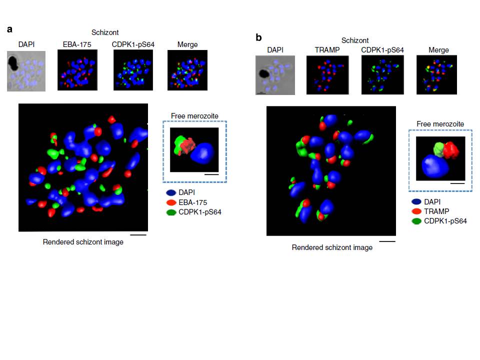
PfCDPK1 phosphorylated at serine-64 is associated with apical parasite structures. (a) A schizont stage parasite stained with DAPI to reveal the nuclei (blue), an antibody to PfEBA-175 to reveal the micronemes (red) and the phospho-specific CDPK1-pS64 antibody (green). A merge of images from all three stains is shown on the far right; this is a representative image from at least three experiments. Also shown is a rendered image where deconvoluted z stacks were reconstructed in 3D, with interpolation. The inset shows a rendered image of a free merozoite from the same preparation . (b) The same as a, but instead of probing with an anti-EBA-175 antibody the preparation was probed with an antibody to the rhoptry
marker PfTRAMP . Scale bars, a-1 mm (insert-0.5 mm), b-1 mm (insert-0.5 mm).
Alam MM, Solyakov L, Bottrill AR, Flueck C, Siddiqui FA, Singh S, Mistry S, Viskaduraki M, Lee K, Hopp CS, Chitnis CE, Doerig C, Moon RW, Green JL, Holder AA, Baker DA, Tobin AB. Phosphoproteomics reveals malaria parasite Protein Kinase G as a signalling hub regulating egress and invasion. Nat Commun. 2015 6:7285
Other associated proteins
| PFID | Formal Annotation |
|---|---|
| PF3D7_0217500 | Calcium-dependent protein kinases; CDPK1 |
| PF3D7_1218000 | thrombospondin-related apical membrane protein |