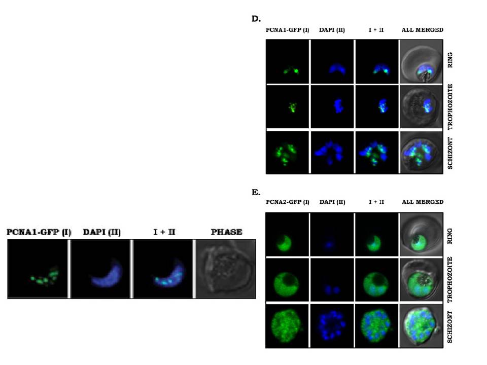
Right panels: Stage-specific expression and subcellular localization of PfPCNA proteins. (D and E) PfPCNA1–GFP and PfPCNA2–GFP transgenic parasite lines were visualized using an Olympus confocal microscope and Olympus Fluoview software. PfPCNA1–GFP (D) predominantly shows nuclear punctate staining co-localized with DAPI. PfPCNA2–GFP (E) shows a diffuse staining pattern across the parasite including the nuclei stained with DAPI. PCNA1–GFP showed distinct nuclear foci similar to endogenous protein (2D). However, these foci were absent from PCNA2–GFP, where a diffuse staining pattern could be seen throughout the parasites with slight enrichment of PCNA2–GFP in the nucleus (E). :pwer left panel: PCNA1 marks the sites of active replication. Live cell imaging of PCNA1-GFP expressing parasites using high resolution confocal microscopy shows distinct PCNA1 foci associated with the nucleus (shown in blue, DAPI) in trophozoite stages of the parasites.
Mitra P, Banu K, Deshmukh AS, Subbarao N, Dhar SK. Functional dissection of proliferating-cell nuclear antigens (1 and 2) in human malarial parasite Plasmodium falciparum: possible involvement in DNA replication and DNA damage response. Biochem J. 2015 470(1):115-29.
Other associated proteins
| PFID | Formal Annotation |
|---|---|
| PF3D7_1226600 | proliferating cell nuclear antigen 2 |