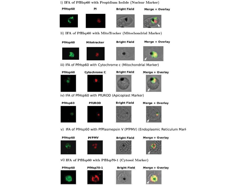
Sub-cellular distribution and localization of PfHsp60 (continued). (A) IFA of localization PfHsp60 in P. falciparum infected erythrocytes with (i) Propidium iodide (Nuclear marker), (ii) Mitotracker, (iii) PfCytochrome C (Mitochondrial marker), (iv) PfUROD (Apicoplast marker), (v) PfPlasmepsin V (PfPMV, endoplasmic reticulum marker), and (vi) PfHsp70-1 (Cytosol marker). PfHsp60 is stained with FITC-conjugated secondary antibody in all parts, while PfUROD, PfPlasmepsin V, PfCytochrome C, and PfHsp70-1 are stained with TRITC-cojugated secondary antibody in (iii), (iv), and (v). In all images, white arrow indicates the erythrocyte membrane. Part (i) shows that PfHsp60 is present only in the extra-nuclear compartment. Parts (ii) and (iii) show that PfHsp60 is present in the cytoplasm as well as in the mitochondria. The yellow signal in the merged panel shows that there is some co-localization. Part (iv) shows a lack of signal overlap with PfUROD in the merge panel. Part (v) shows a lack of signal overlap with PfPlasmepsin V in the merge panel, which rules out its localization in endoplasmic reticulum. Part (vi) shows that there is co-localization between the cytosolic protein PfHsp70-1 and PfHsp60, which indicates that PfHsp60 indeed accumulates in the cytosol
Padma Priya P, Grover M, Tatu US, Natarajan V. Characterization of Precursor PfHsp60 in Plasmodium falciparum Cytosol during Its Asexual Development in Human Erythrocytes. PLoS One. 2015 Aug 28;10(8):e0136401. PMID:
Other associated proteins
| PFID | Formal Annotation |
|---|---|
| PF3D7_0818900 | PfHsp70-1 |
| PF3D7_1015600 | heat shock protein 60 |
| PF3D7_1323500 | PEXEL protease plasmepsin V |
| PF3D7_1404100 | cytochrome c, putative |