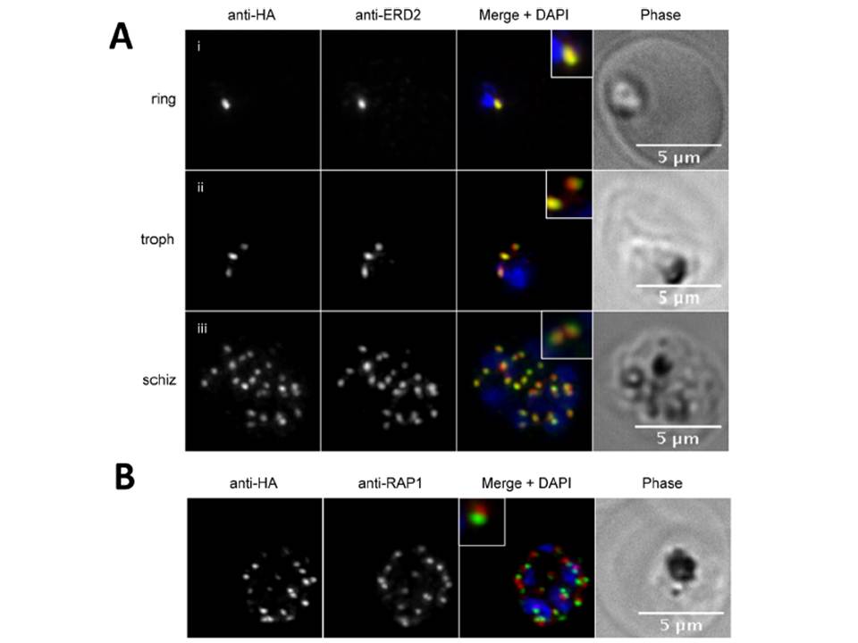
PfPRP2-RFA overlaps extensively with the Golgi apparatus throughout the erythrocytic cycle. (A) In ring stages, PfPRP2-RFA is found as a single punctate pattern colocalizing with the cis-Golgi marker ERD2 (Ai). In trophozoites, prior to nuclear division, the pattern associated with both PfPRP2-RFA and ERD2 becomes triple dots, showing the division of the Golgi, prior to nuclear replication (Aii). In schizont stages, the
multiple PfPRP2-RFA signals still colocalize with the cis-Golgi protein ERD2 (Aiii). (B) In mature schizonts, PfPRP2-RFA is found in close proximity but does not overlap with the rhoptry marker RAP1. RAP1, rhoptry associated protein 1. Nuclei of parasites were stained with DAPI (blue). The fluorescence of PfPRP2-RFA is pseudocolored in green and other markers are in red. Scale bar represents 5μm.
Hallée S, Richard D. Evidence that the Malaria Parasite Plasmodium falciparum Putative Rhoptry Protein 2 Localizes to the Golgi Apparatus throughout the Erythrocytic Cycle. PLoS One. 2015 Sep 16;10(9):e0138626.
Other associated proteins
| PFID | Formal Annotation |
|---|---|
| PF3D7_1320000 | rhoptry protein 2, putative golgi protein 1 |
| PF3D7_1353600 | ER lumen protein retaining receptor |