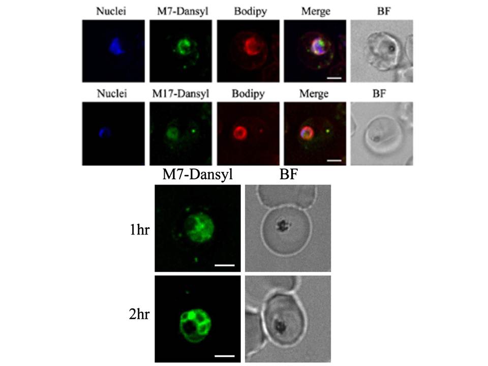
Upper panel: Location of Dansyl-MAS 7 and MAS 17 peptides, within the parasite infected erythrocyte. Parasite nuclei are shown in blue, Dansyl fluorescence in green and BODIPY-TR-Ceramide in red. A merge and bright field (BF) image are shown. Scale bar = 3 μm. MAS-7 probably binds to adenylyl cyclase .
Lower panel: Location of Dansyl-MAS 7 peptide, within the parasite infected erythrocyte following 1 hour (upper panel) and 2 hour (lower panel) incubation. Scale bar = 3 μm.
Peatey CL, Dixon MW, Gardiner DL, Trenholme KR. Temporal evaluation of
commitment to sexual development in Plasmodium falciparum. Malar J. 2013 12:134.
Other associated proteins
| PFID | Formal Annotation |
|---|---|
| PF3D7_1404600 | Adenylyl cyclase Aca |