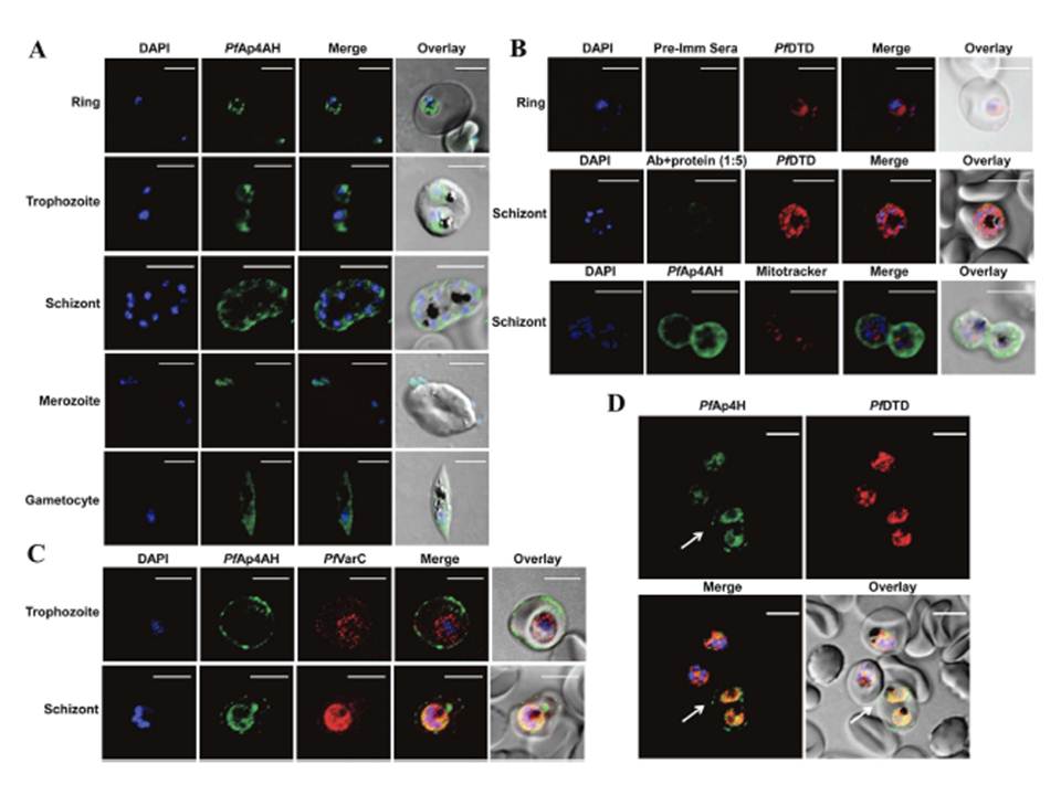
Spatial distribution of P. falciparum Ap4AH during erythrocytic schizogony. Shown are DAPI staining of nucleus in blue and PfAp4AH stained with Alexa 488 in green. (A) Confocal microscopy-data based spatial distribution of PfAp4AH in infected RBCs. PfAp4AH is non-nuclear in blood stages of the parasite and resides in its cytoplasm. (B) Non-mitochondrial localization with various controls is shown. Upper panel shows pre-immune serum (Pre-Imm Sera) control which does not stain the parasite or RBCs. Middle panel shows competitive binding of anti-PfAp4AH antibody to infected cells, where anti-PfAp4AH antibodies were incubated with recombinant PfAp4AH protein in 1:5 ratio. Pf D-tyrosyl-tRNATyr deacylase (DTD) is a cytoplasmic marker. Lower panel shows non-mitochondrial localization where mtochondria are stained in red. (C) RBC membrane localization of PfAp4AH during trophozoite and schizont stages of parasite. VarC is a marker for infected RBC membrane localization. (D) A field view of anti-PfAp4AH antibody staining of infected RBCs. Significant fraction of cells (~50%) showed membrane localization of PfAp4AH - here cell is marked with white arrow. Uninfected RBCs (without DAPI and PfDTD staining here) are unstained. White scale bar in confocal figures is of 5 μm
Sharma A, Yogavel M, Sharma A. Structural and functional attributes of malaria parasite diadenosine tetraphosphate hydrolase. Sci Rep. 2016 Feb 1;6:19981.
Other associated proteins
| PFID | Formal Annotation |
|---|---|
| PF3D7_0520600 | bis(5'-nucleosyl)-tetraphosphatase [asymmetrical] Ap4A hydrolase |
| PF3D7_1108200 | D-tyrosyl-tRNA(Tyr) deacylase |