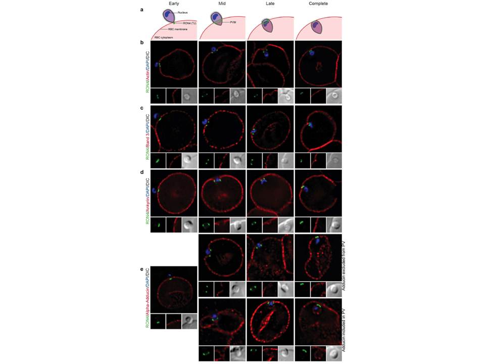
Imaging of the erythrocyte cytoskeleton throughout merozoite invasion. Widefield deconvolution fluorescence imaging of fixed merozoite invasion samples labelled with anti-RON4, DAPI and markers of erythrocyte cytoskeletal proteins. (a) Schematic showing how RON4 labelling allows discernment of early, mid, late and complete invasion events. Early in invasion, during apical attachment, RON4 appears as a single point of fluorescence before expanding to form a ring around the invading merozoite as invasion progresses. When imaged in two dimensions, this ring appears as two single points of fluorescence on either side of the merozoite. The right junction marks the boundary between the erythrocyte plasma membrane and the parasitophorous vacuole membrane, and closes at the base of the parasite at the end of invasion. (b) Erythrocyte actin, labelled by phalloidin-Alexa594. (c,d) Band 3, ankyrin and adducin in invasion labelled with specific antibodies. PVM, parasitophorous vacuole membrane; RBC, red blood cell; TJ, tight junction
Zuccala ES, Satchwell TJ, Angrisano F, Tan YH, Wilson MC, Heesom KJ, Baum J.
Quantitative phospho-proteomics reveals the Plasmodium merozoite triggers
pre-invasion host kinase modification of the red cell cytoskeleton. Sci Rep. 2016
Feb 2;6:19766. doi: 10.1038/srep19766.