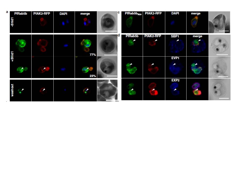
PfRab5b expression disturbed the transport of PfAK2 to the TVN. a In the absence of Shld1, PfAK2-RFP (red) localized to the TVN, while PfRab5b-YFP-DD signal was not detect (−Shld1, upper panel). After Shld1 stabilization for 48 h, PfRab5b-YFP-DD (green) and PfAK2-RFP co-localized in the TVN-like structures (+Shld1, middle-upper panel). In 77 % of PfRab5b-YFP-DD-positive parasites, PfAK2-RFP localized to the TVN, while in 23 % of PfRab5b-YFP-DD positive parasites, PfAK2-RFP signals also accumulated in the punctate compartment within the parasite (+Shld1, middle-lower
panel, arrowhead). Co-localization of PfRab5b-YFP-DD and PfAK2-RFP at the punctate compartment remained even after removal of Shld1 (washout, lower panel, arrowhead). c No punctate localization of PfAK2-RFP was apparent with co-expression of PfRab5bN20-YFP-DD. d Effect of PfRab5b over-expression on the exported proteins PfSBP1, PfEVP1, and PfEXP2. In contrast to PfAK2-RFP, these proteins did not accumulate in the PfRab5b- and PfAK2-double positive punctate compartments (arrowhead).
Ebine K, Hirai M, Sakaguchi M, Yahata K, Kaneko O, Saito-Nakano Y. Plasmodium Rab5b is secreted to the cytoplasmic face of the tubovesicular network in infected red blood cells together with N-acylated adenylate kinase 2. Malar J. 2016 15:323.
Other associated proteins
| PFID | Formal Annotation |
|---|---|
| PF3D7_1310600 | ras-related protein Rab-5B secretory complex protein 61 alpha |