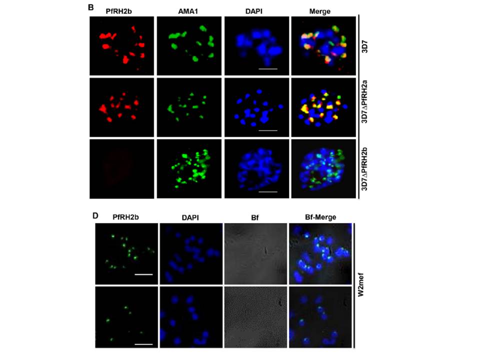
B. IFA indicates the specificity of mAb A7 in schizont stage parasites. PfRH2b (PfRH2b, red) co-stained with AMA1 (AMA1, green), DAPI for nuclei (blue) in 3D7, 3D7DPfRH2a and 3D7DPfRH2b schizont. In the merged images, areas of overlap between the red and the green signals are shown in yellow. Scale bar indicated in white represents 2 mm. D. Non-permeabilized W2mef merozoites show surface expression of PfRH2b using mAb A7. PfRH2b shown green (RH2b A7), DAPI for nuclei (blue) and bright field (Bf) images shown in gray. Scale bar indicates 2 mm. (3D7DPfRH2a (clone in which PfRH2a has been deleted), 3D7DPfRH2b (clone in which the PfRH2b gene has been deleted) parasites. The punctate staining pattern observed for A7 was in close proximity to the staining observed with an antibody targeting the micronemal protein Apical membrane antigen 1 (AMA1), indicating an apical location of PfRH2b within the merozoite.
Aniweh Y, Gao X, Gunalan K, Preiser PR. PfRH2b specific monoclonal antibodies inhibit merozoite invasion. Mol Microbiol. 2016 Nov;102(3):386-404.
Other associated proteins
| PFID | Formal Annotation |
|---|---|
| PF3D7_1133400 | apical membrane antigen 1 |
| PF3D7_1335300 | reticulocyte binding protein 2 homologue b, ALKBH5 |