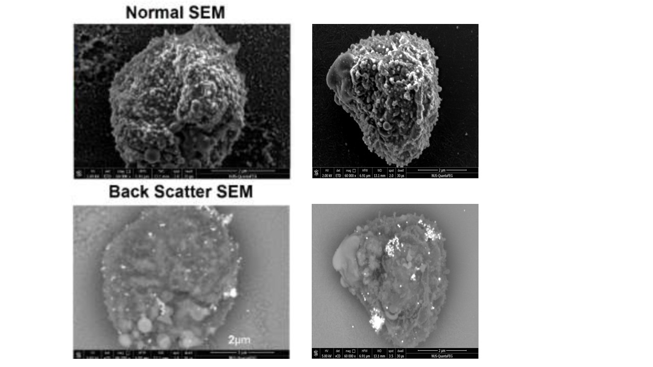
Clustering of surface exposed STEVOR in proximity to knobs. Normal and backscatter SEM images of a typical late stage iRBC from 5A clone. Normal image shows the surface topology, while the backscatter image shows the bright 10nm gold nanoparticles. Scale bar, 2μm. STEVOR molecules on the cell surface were immunostained with anti-S1 serum followed by secondary antibodies conjugated with 10nm gold (Au) nanoparticles These
images revealed that STEVOR were distributed on the surface in various cluster sizes.
Singh H, Madnani K, Lim YB, Cao J, Preiser PR, Lim CT. Expression dynamics & physiologically relevant functional study of STEVOR in asexual stages of Plasmodium falciparum infection. Cell Microbiol. 2016 Dec 28.