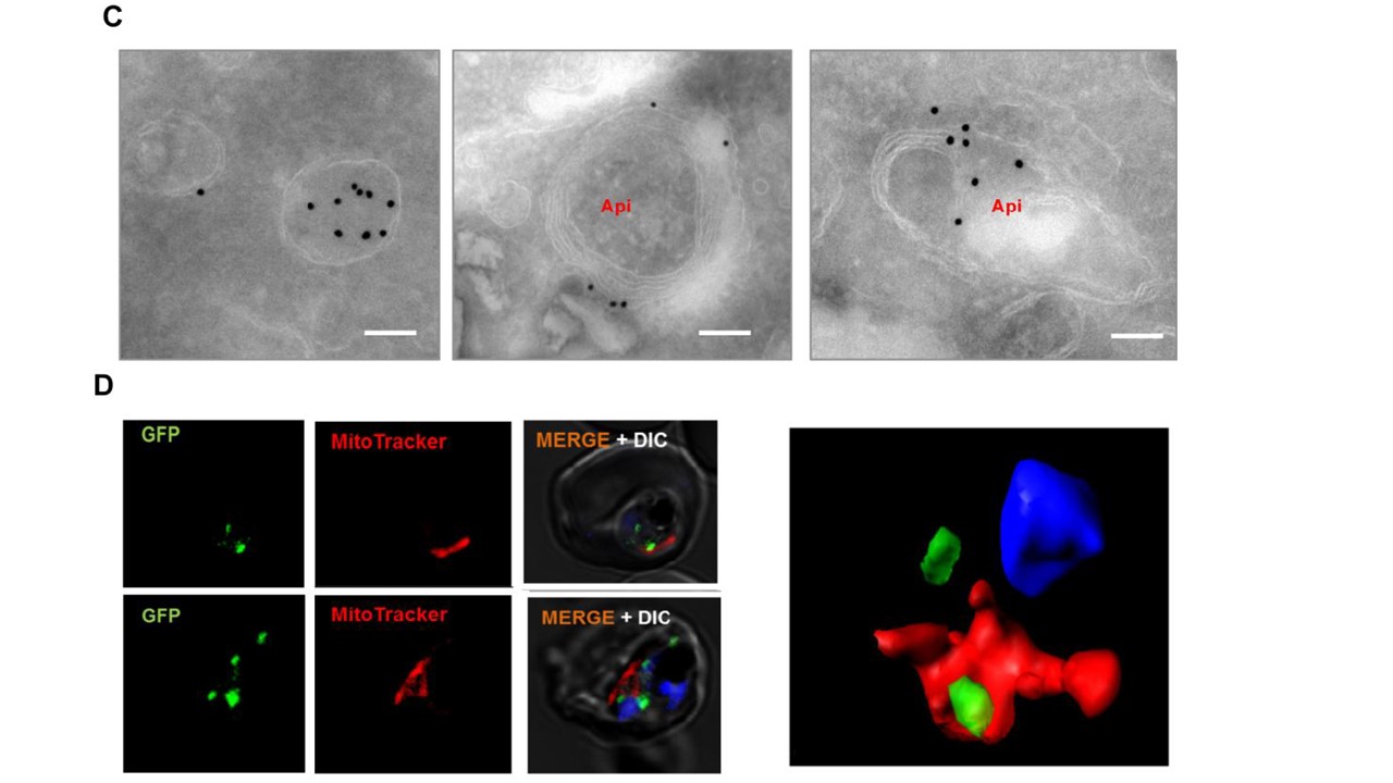
Sub-cellular localisation studies of PfOTU in transgenic parasites. (C) Localisation of PfOTU by immune-electron microscopy. Ultra-thin sections of transgenic PfOTU-RFA parasites were labelled with anti-GFP antibody and gold-labelled secondary antibody. The labelling was seen in the cytosolic vesicles or associated with the apicoplast having characteristic four membranes. Scale bar-100 nm. In trophozoite stage parasites, the gold labelling was observed in membrane bound vesicles of 100-200nm diameter. In some sections these labelled vesicles were present near the apicoplast, while in some cases the immuno-staining was observed at the apicoplast surface overlapping with its membrane. (D) Confocal microscopic images showing the mitoTracker staining (red) in PfOTU-RFA transgenic parasites; corresponding three dimensional reconstruction using series of Z-stack images showing GFP labelled vesicles (green) distinct from the mitochondria (red).vStaining of transgenic parasites with MitoTacker Red CMXRos showed elongated/branched mitochondria whereas the GFP-labelled vesicles were present distinct from this organelle,
Datta G, Hossain ME, Asad M, Rathore S, Mohmmed A. Plasmodium falciparum OTU-like cysteine protease (PfOTU) is essential for apicoplast homeostasis and associates with non-canonical role of Atg8. Cell Microbiol. 2017 Apr 18. [Epub ahead of print]