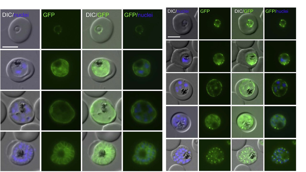
Proteins with TM that are located in the PV. Live cell microscopy images of the endogenously GFP-tagged alkaline phosphatase and EXP3. Left panel: a young and an old schizont are shown to illustrate the change from a staining surrounding the bulk of the still forming merozoites (third row of images) to a ‘bunch of grape’ type pattern where each merozoite is surrounded by GFP signal after the PPM is invaginated (bottom row of images). Right panel: a late trophozoite (third row of images), a young schizont (fourth row of images) and a late schizont (bottom row of images) are shown to demonstrate the change from a peripheral to a parasite internal staining .
Khosh-Naucke M, Becker J, Mesén-Ramírez P, Kiani P, Birnbaum J, Fröhlke U, Jonscher E, Schlüter H, Spielmann T. Identification of novel parasitophorous vacuole proteins in P. falciparum parasites using BioID. Int J Med Microbiol. 2017 Jul 27. [Epub ahead of print]
Other associated proteins
| PFID | Formal Annotation |
|---|---|
| PF3D7_1024800 | exported protein 3 |