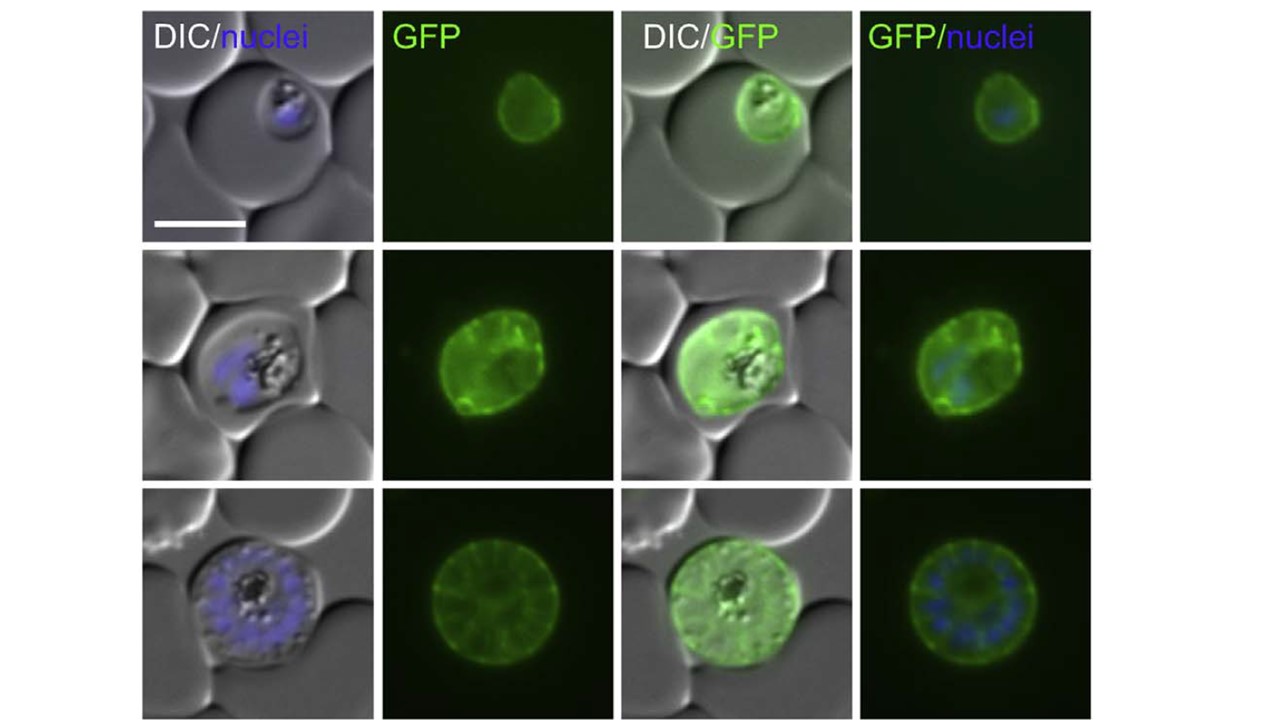
Proteins without TM that are located in the PV. The top microscopy panel row shows a ring and a trophozoite stage, the middle row shows a schizont and the bottom row shows a late schizont. Inspection of late stage parasites - in this phase, the PPM is
internalised to surround the merozoites, leading to a ‘bunch of grape’ - no typical bunch of grape staining pattern was observed. Instead the signal surrounded the merozoites as a group, not individually.
Khosh-Naucke M, Becker J, Mesén-Ramírez P, Kiani P, Birnbaum J, Fröhlke U, Jonscher E, Schlüter H, Spielmann T. Identification of novel parasitophorous vacuole proteins in P. falciparum parasites using BioID. Int J Med Microbiol. 2017 Jul 27. [Epub ahead of print]