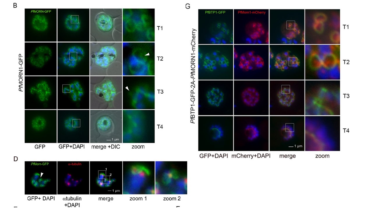
Colocalization of BTP1 with MORN1. (B) Localization of P. falciparum (pf)MORN1–GFP in unfixed parasites. MORN1–GFP reveals a contractile ring structure that moves along the longitudinal axes of the nascent merozoite (T1–T4) with an additional punctate structure that might represent the centrocone (marked with arrowheads). Nuclei were stained with DAPI (blue). Enlargement of selected areas are marked with a white square and referred to as ‘zoom’ (a fourfold magnification). (D) Colocalization of MORN1–GFP with α-tubulin (red) that highlights the microtubule-organizing centers (MTOCs) at this stage. Although the MORN1-marked ring structure is clearly distinct from the MTOC (zoom 1), there is a close association of the MTOC with the additional MORN1-marked structure (zoom 2). (G) Colocalization analysis of BTP1–GFP (green) with MORN1–mCherry (red) revealed their identical localization during schizogony, except for the exclusively MORN1–mCherry-highlighted putative centrocone.
Kono M, Heincke D, Wilcke L, Wong TW, Bruns C, Herrmann S, Spielmann T, Gilberger TW. Pellicle formation in the malaria parasite. J Cell Sci. 2016 129(4):673-80. PMID: 26763910
Other associated proteins
| PFID | Formal Annotation |
|---|---|
| PF3D7_0611600 | basal complex transmembrane protein 1 |