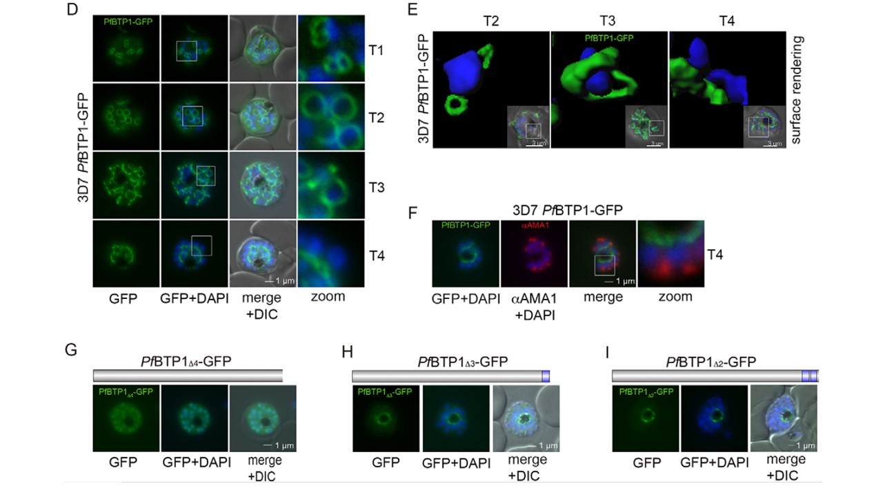
Expression and localization of BTP1. (D) BTP1–GFP can be visualized as a contractile ring structure in unfixed late-stage parasites (T1–T4). It first appears at the apical pole of the nascent merozoite (T1) and ends up at the basal pole of the mature merozoite (T4). Nuclei were stained with DAPI (blue). Enlargement of selected areas are marked with a white square and referred to as ‘zoom’ (a fourfold magnification). (E) Surface-rendering plot of an indicated section, highlighting the spatial relationship between nuclei and the basal-complex ring. (F) Colocalization of BTP1–GFP and the apical membrane antigen 1 (αAMA1; red); AMA1 localizes in the micronomes at the apical pole opposing BPT1, confirming the basal localization of BTP1–GFP at the end of daughter cell formation. (G–I) Localization of the BPT1 deletion mutants. (G) Deletion of all four transmembrane domains leads to a nuclear and cytosolic distribution of BTP1Δ4−GFP. (H) Expression of BTP1 with one (BTP1Δ3−GFP) or two (BTP1Δ2−GFP, I) transmembrane domains results in localization of mutant BTP1 at the food vacuole membrane. DIC, differential interference contrast.
Kono M, Heincke D, Wilcke L, Wong TW, Bruns C, Herrmann S, Spielmann T, Gilberger TW. Pellicle formation in the malaria parasite. J Cell Sci. 2016 129(4):673-80.
Other associated proteins
| PFID | Formal Annotation |
|---|---|
| PF3D7_1133400 | apical membrane antigen 1 |