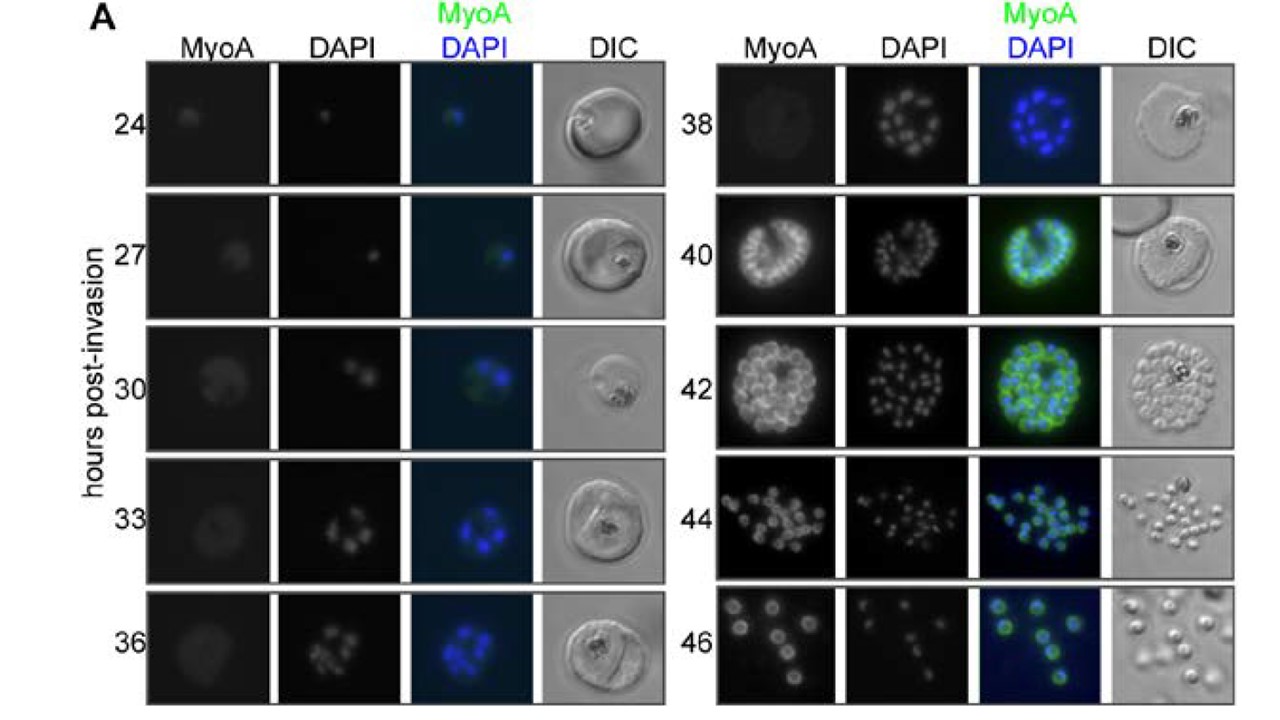
MyoA-GFP expression and glideosome complex formation during the late stages of intracellular development of P. falciparum in the red blood cell. (A) MyoA-GFP expression was detected by live fluorescence microscopy and the nuclei were detected by staining with DAPI. The merged colour image with MyoA-GFP (green) and DAPI (blue) and differential interference contrast (DIC) bright field images are also shown. Schizogony starts at around 30 hours post-invasion and MyoA-GFP is detected from 38–40 hours in multinucleated forms. Scale bar: 2 µm. MyoA-GFP was largely undetectable by live fluorescence microscopy until approximately 38 hours after merozoite invasion of an erythrocyte, but was present in later stages at the periphery of developing segmented schizonts and free merozoites (but not associated with the food vacuole/residual body), consistent with its proposed location at the IMC (Fig. 2A). No GFP signal was detected in ring or trophozoite stages indicating that the protein is not synthesised at this time and any MyoA carried through with the invading merozoite is degraded rapidly following erythrocyte invasion.
Green JL, Wall RJ, Vahokoski J, Yusuf NA, Ridzuan MAM, Stanway RR, Stock J, Knuepfer E, Brady D, Martin SR, Howell SA, Pires IP, Moon RW, Molloy JE, Kursula I, Tewari R, Holder AA. Compositional and expression analyses of the glideosome during the Plasmodium life cycle reveal an additional myosin light chain required for maximum motility. J Biol Chem. 2017 Sep 11.