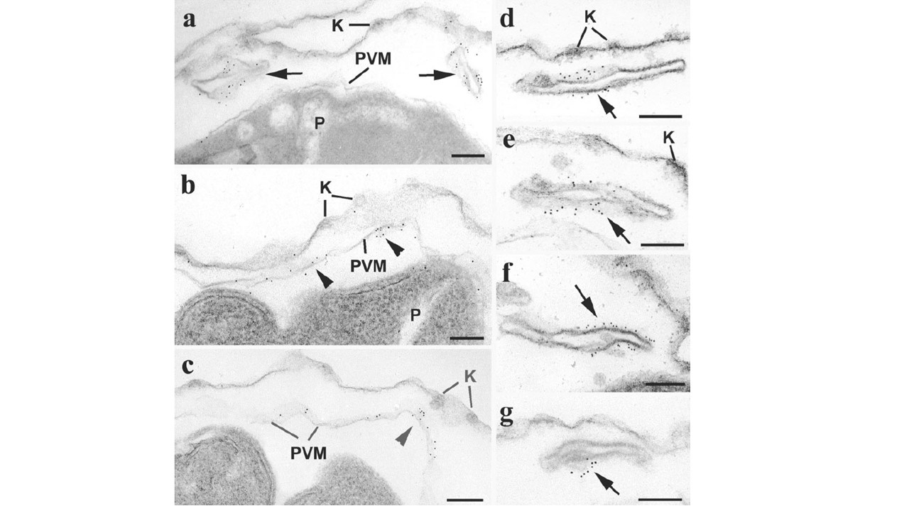
Transmission electron microscopy of immunogold labeling of MAHRP1 in saponin-permeabilized parasitized RBCs. (a to c) Sections through the periphery of infected RBCs showing labeling of the Maurer’s clefts (arrows) with variable labeling of the PV membrane (PVM; arrowheads). In most sections (a), the PVM is unlabeled; however, in a few sections (b and c), there is focal labeling. (d to g) Details of Maurer’s clefts showing that the gold particles are mainly located in the central region (arrows). K, knobs; P, parasite. Bars, 100 nm.
Spycher C, Rug M, Klonis N, Ferguson DJ, Cowman AF, Beck HP, Tilley L. Genesis of and trafficking to the Maurer's clefts of Plasmodium falciparum-infected erythrocytes. Mol Cell Biol. 2006 26(11):4074-85.