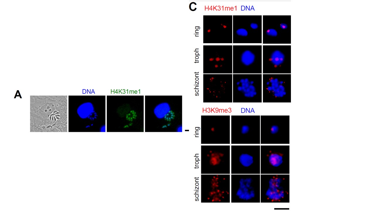
Distribution of H4K31me1 in parasites nuclei (a) Immunofluorescence analysis of H4K31me1 (in green) in intracellular parasite nuclei. DNA was stained with Hoechst (blue). Scale bar, 10 mm. In situ, the H4K31me1 modification appeared uniformly distributed within the nucleus of dividing parasites but surprisingly no signal was detected in the nucleus of the infected human cell. (c) Immunofluorescence analysis of H4K31me1 (in red) or H3K9me3 in asexual stages of Pf-3D7. Scale bar, 5 m. Data are representative of three independent experiments. H4K31me1 displayed a peculiar condensed punctate pattern (c), similar to the H3K9me3 mark (Lopez-Rubio et al., 2009) at the nuclear periphery,
which is reminiscent of heterochromatin/subtelomeric regions clustering (Freitas-Junior et al., 2000). P. falciparum centromeres also clustered prior to and throughout mitosis and cytokinesis leading to single nuclear location from early trophozoites to mature schizonts (Hoeijmakers et al., 2012). Therefore, H4K31me1-containing foci could be associated with subtelomeric or/and centromeric regions.
Sindikubwabo F, Ding S, Hussain T, Ortet P, Barakat M, Baumgarten S, Cannella D, Palencia A, Bougdour A, Belmudes L, Couté Y, Tardieux I, Botté CY, Scherf A, Hakimi MA. Modifications at K31 on the lateral surface of histone H4 contribute to genome structure and expression in apicomplexan parasites. Elife. 2017 Nov 4;6. pii: e29391.
Other associated proteins
| PFID | Formal Annotation |
|---|---|
| PF3D7_1105000 | histone H4 |