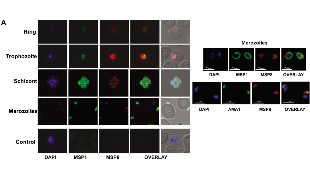
(A) Localization of PfMSP8 in blood stages of the parasite. PfMSP8 localization was evaluated by IFA using acetone-methanol-fixed slides of asynchronous P. falciparum (FVO)-infected erythrocytes. The smears were costained with rabbit anti-rPfMSP8 IgG and mouse anti-PfMSP119 (FVO) monoclonal antibody 5.2. Bound IgGs were detected by TRITC-conjugated goat anti-rabbit IgG and FITC-conjugated goat anti-mouse IgG and parasite DNA stained with DAPI. Shown are representative images of parasite DNA-specific fluorescence (blue) (column 1), PfMSP1-specific fluorescence (green) (column 2), and PfMSP8-specific fluorescence (red) (column 3), merged images of PfMSP1 and PfMSP8 (column 4), and merged images of DAPI, PfMSP1, PfMSP8, and the corresponding DIC images (column 5). Representative images of ring-, trophozoite-, schizont-, and merozoite-stage parasites are shown. “Control” represents corresponding images of infected RBCs stained with normal mouse sera and adjuvant control rabbit IgG. (B) Merozoite-associated PfMSP8. In free merozoites, colocalization of PfMSP8 with either PfMSP1 (top) or PfAMA1(bottom) was evaluated by IFA as described above using rabbit anti-rPfMSP8 IgG,MAb5.2 (mouse anti-PfMSP119 FVO), and MAb 4G2 (rat anti-PfAMA1 FVO) with images acquired by confocal microscopy.
Alaro JR, Angov E, Lopez AM, Zhou H, Long CA, Burns JM Jr. Evaluation of the immunogenicity and vaccine potential of recombinant Plasmodium falciparum merozoite surface protein 8. Infect Immun. 2012 80(7):2473-84
Other associated proteins
| PFID | Formal Annotation |
|---|---|
| PF3D7_0502400 | ring-stage membrane protein 1 merozoite surface protein 8 |
| PF3D7_1133400 | apical membrane antigen 1 |