PfClpC-DDD parasites (1G8 clone) grown for 10 days without TMP and supplemented with IPP. These PfClpC-DDD parasites were stained with
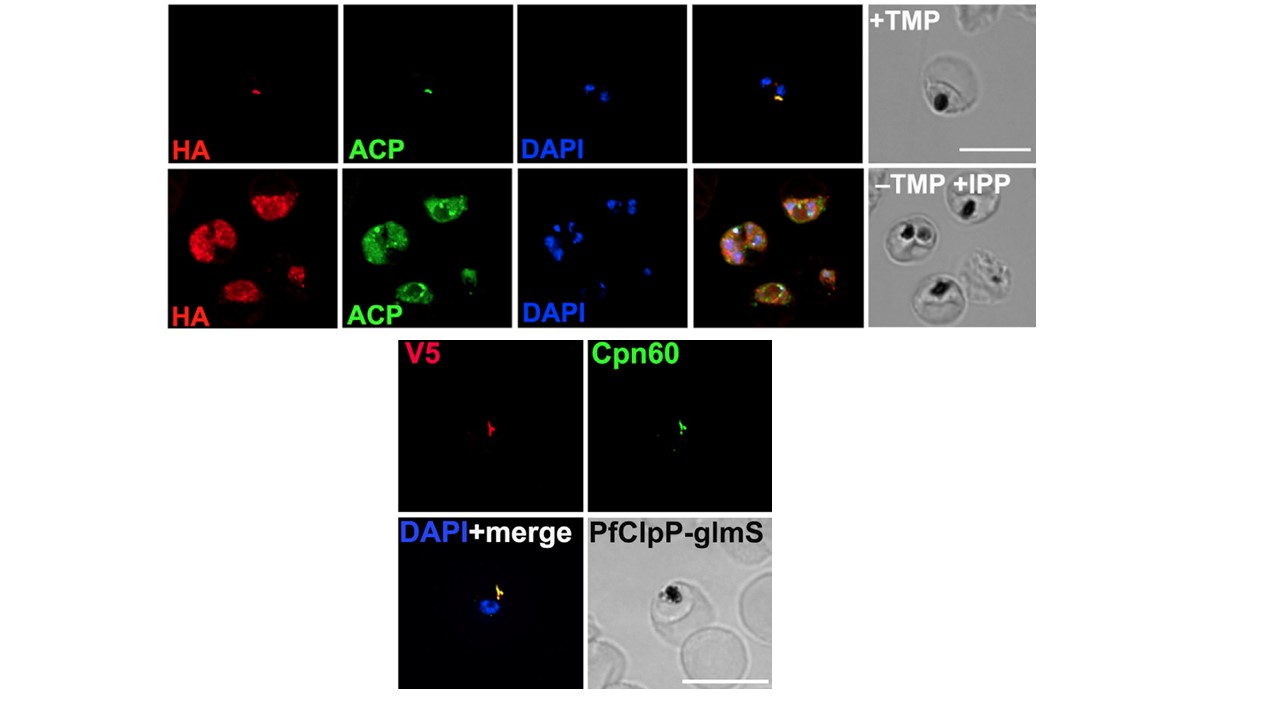
specific antibodies. Images from left to right are anti-HA (red), anti-ACP (green), DAPI (parasite nucleus, blue), fluorescence merge, and phase. Z stack images were deconvolved and projected as a combined single image. Scale bar, 5 mm. Immunofluorescence microscopy revealed, however, that IPP-treated PfClpC-DDD parasites survived in the absence of an apicoplast and retained the vesicle-like structures containing PfClpC and ACP.
Lower panel. Immunofluorescence imaging of unsynchronized PfClpP-glmS1 parasites stained with specific antibodies. Images from left to right are anti-V5 (red) and anti-Cpn60 (green) in the upper panel and DAPI (parasite nucleus, blue), along with fluorescence merge, and phase in the lower panel. Z stack images were deconvolved and projected as a combined single image. Scale bar, 5 mm. We were able to detect the apicoplast localization of PfClpP by staining with anti-V5 and anti-Cpn60.
Florentin A, Cobb DW, Fishburn JD, Cipriano MJ, Kim PS, Fierro MA, Striepen B, Muralidharan V. PfClpC Is an Essential Clp Chaperone Required for Plastid Integrity and Clp Protease Stability in Plasmodium falciparum. Cell Rep. 2017 21(7):1746-1756.
Other associated proteins
| PFID | Formal Annotation |
|---|---|
| PF3D7_1232100 | 60 kDa chaperonin |
| PF3D7_1406600 | ATP-dependent Clp protease regulatory subunit Clpc, putative |