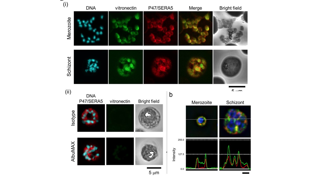
Localization of VTN and P47 domain/SERA5. (a) (i) Representative immunofluorescence assay (IFA) images of merozoites and schizont. (ii) Upper panels, Representative IFA images of schizont. VTN was probed with an isotype control mAb instead of anti-VTN mAb. Lower panels, Representative IFA images of schizont cultured in AlbuMAX-I medium. Target proteins were probed with anti-VTN mAb (green), anti-SE36 rabbit serum (red), and DAPI nuclear stain (blue). Scale bar, 5 μm. (b) Representative IFA images of merozoite and schizont stages under deconvolution microscopy. Probes used were the same as in panel a. Upper panels show the localization of P47 domain/SERA5 during merozoite and schizont stages. Lower panels show the intensity of green and red signals at the section of the gray line in the upper panel. Scale bar, 1 μm.
Tougan T, Edula JR, Takashima E, Morita M, Shinohara M, Shinohara A, Tsuboi T, Horii T. Molecular Camouflage of Plasmodium falciparum Merozoites by Binding of Host Vitronectin to P47 Fragment of SERA5. Sci Rep. 2018 Mar 22;8(1):5052.