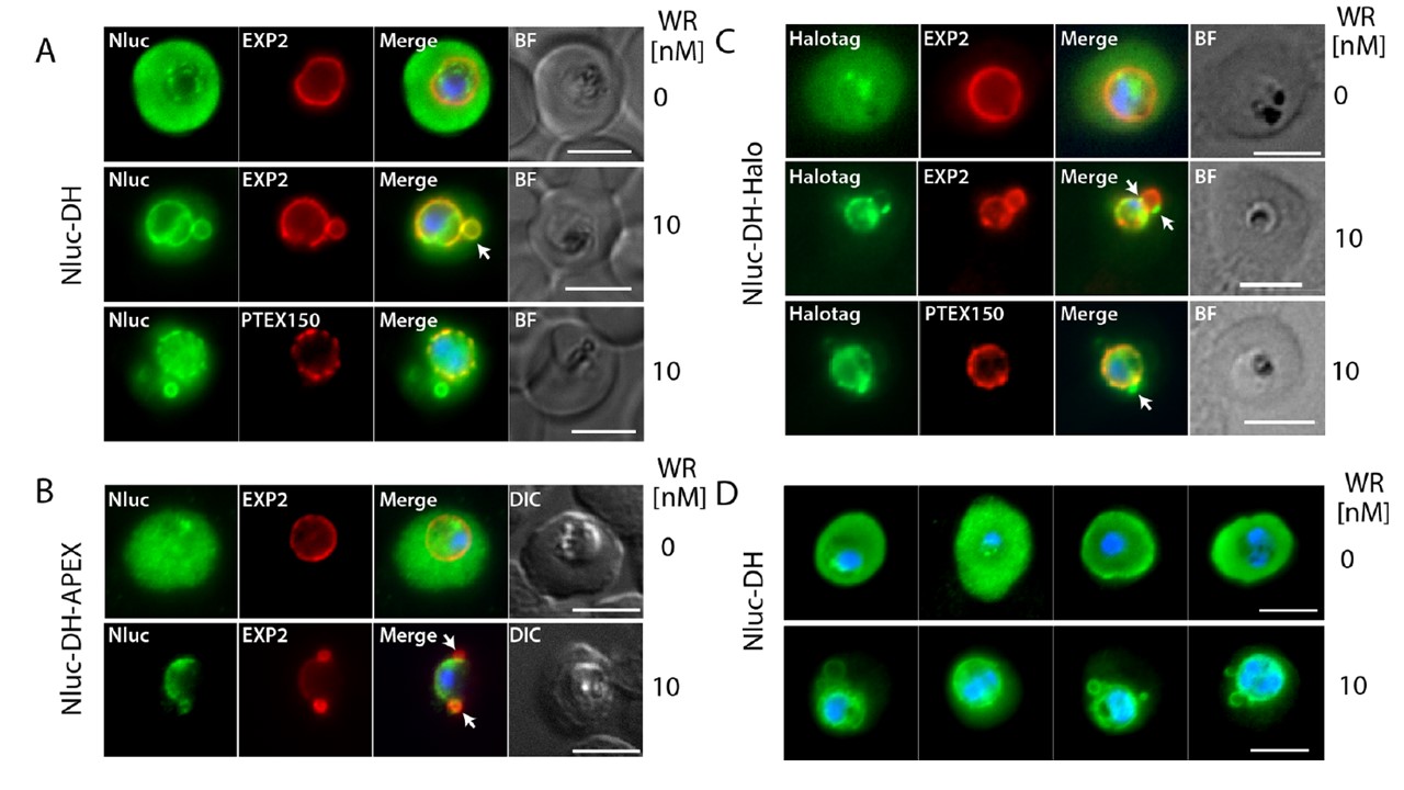
Widefield immunofluorescence microscopy of Nluc-DH, Nluc-DH-APEX and Nluc-DH-Halo trophozoites (parasites, expressing exported nanoluciferase) reveals treatment with WR99210 blocks protein export into the erythrocyte compartment. (A-C) Parasite lines imaged are indicated on the left, antibody probes are shown at the top of the image panels and the
concentration of WR used is on the right. For Nluc-DH-Halo the Halotag was stained with the Oregon green dye. White arrows indicate the EXP2 and trapped reporter protein loops appearing to project from the parasitophorous vacuole membrane. (D) A selection of Nluc-DH parasites treated -/+ WR showing Nluc (green) trapped within the parasite especially around the nuclei (blue). Scale bar = 5 μm. DIC, Differential interference contrast; BF, Brightfield. in WR-treated trophozoites EXP2 often labelled loop structures attached to the PVM that were also partly labelled for all the Nluc-DH reporters (A-C, white arrows).
To determine if these loops contained the PTEX150, Nluc-DH and Nluc-DH-Halo trophozoites were probed with anti-HA IgG to detect PTEX150-HA and this indicated its near absence from the loops.
Charnaud SC, Jonsdottir TK, Sanders PR, Bullen HE, Dickerman BK, Kouskousis B, Palmer CS, Pietrzak HM, Laumaea AE, Erazo AB, McHugh E, Tilley L, Crabb BS, Gilson PR. Spatial organisation of protein export in malaria parasite blood
stages. Traffic. 2018 Apr 26. [Epub ahead of print]
Other associated proteins
| PFID | Formal Annotation |
|---|---|
| PF3D7_1436300 | translocon component PTEX150 |