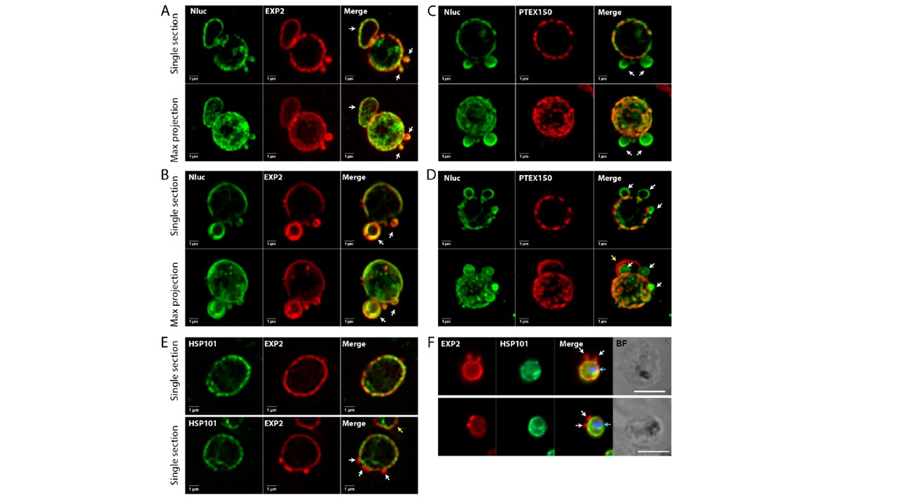
Structured illumination microscopy of WR-treated Nluc-DH parasites (expressing exported nanoluciferase) trophozoites showing trapped cargo proteins localising with PTEX proteins in the PVM. (A, B) (Top) Single z optical sections of parasites stained with anti-Nluc and anti-EXP2 IgGs showing both proteins colocalised to PV puncta and loops projecting from the PVM that are indicated with white arrows. (Bottom) Maximum intensity projections of serial optical z sections showing both proteins colocalised into punctate regions on the parasite surface. (C, D) (Top) Single sections of Nluc-DH parasites stained with anti-Nluc and anti-PTEX150-HA IgGs showing both proteins localize to PV puncta but PTEX150 does not colocalise to the surface loops. (Bottom) maximum intensity projections showing both proteins localise to surface micro-domains. Yellow arrow in maximum projection of (D) indicates second parasite co-infecting the erythrocyte. Scale bars = 1 μm. (E, F) Respective SIM and widefield images of HSP101-HA trophozoites not transfected with Nluc reporters probed for EXP2 and HSP101 via HA monoclonal IgG. Arrows indicate small EXP2 positive PVM loop ‘buds’ that do not stain for HSP101. Blue arrows in (F) indicate perinuclear HSP101 labelling. Scale bars for SIM (E) and widefield (F) images are 1 and 5 μM, respectively.
Charnaud SC, Jonsdottir TK, Sanders PR, Bullen HE, Dickerman BK, Kouskousis B, Palmer CS, Pietrzak HM, Laumaea AE, Erazo AB, McHugh E, Tilley L, Crabb BS, Gilson PR. Spatial organisation of protein export in malaria parasite blood stages. Traffic. 2018 Apr 26. [Epub ahead of print]
Other associated proteins
| PFID | Formal Annotation |
|---|---|
| PF3D7_1436300 | translocon component PTEX150 |
| PF3D7_1471100 | exported protein 2 |