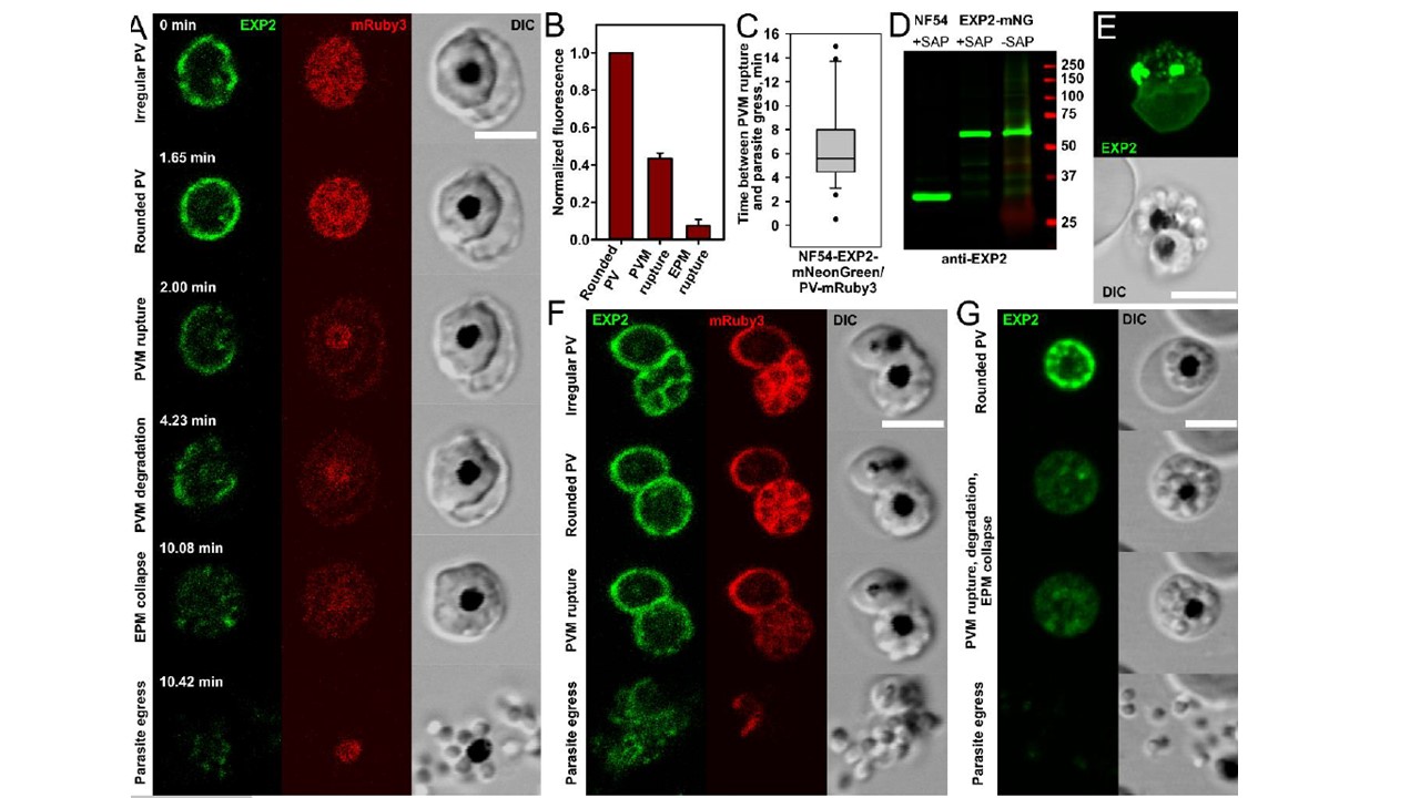
Rupture and degradation of vacuolar membrane and the following steps of erythrocyte membrane transformation. A. Morphologically distinct stages of parasite egress illustrated by a representative recording of parasite egress: the irregular vacuole undergoes transformation into the rounded vacuole and then suddenly ruptures and progressively degrades. This step is associated with redistribution of the vacuolar content to the entire erythrocyte (the first drop of mRuby3 fluorescence) and initiation of the gradual reshaping of the erythrocyte membrane until it collapses on the schizont body. Parasite egress (the disappearance of mRuby3 signal and dispersion of parasites) ends the replicative cycle. B. The drop of mRuby3 fluorescence (PV soluble protein) after sequential rupture of PVM and EPM. The mean fluorescence of all frames between PVM and EPM rupture (the second column) and after EPM rupture (the third column) was normalized to the mean level of fluorescence during the rounded vacuole stage (the first column); (Mean+SEM, n=12). C. Estimated time between PVM rupture and parasite egress (n=27, double labeled parasites, time-lapse recordings of parasite egress). D. Immunoblot detecting EXP2 in asynchronous NF54 parent and EXP2-mNeonGreen purified schizonts shows preservation of the fusion protein in late infected schizonts. Predicted molecular mass after signal peptide cleavage: EXP2: 30.8 kDa; EXP2-mNeonGreen: 57.8 kDa. Comparison of saponin treated (to permeabilizes the EPM and PVM without solubilizing membrane proteins) and untreated samples was performed for two purposes: 1) to improve protein labeling by releasing erythrocyte hemoglobin and 2) to assess the possibility that soluble EXP2 is accumulated in membrane-bound compartments. E. Difference between the maturation times of two parasites within one erythrocyte was exploited to show that rupture and degradation of one vacuole does not affect the integrity of another vacuole. The maximum intensity projection of AiryScan Z stack images of a doubly-infected erythrocyte. Green color: Exp2-mNeonGreen. Note a diffused signal of fluorescence from the PVM surrounded trophozoite (lower cell) and fragmented PVM in the upper schizont. F. Rupture of one vacuole (lower right schizont) in the doubly-infected cells does not affect permeability of the second PVM (upper left trophozoite preserves mRuby3 inside the vacuole), but parasite egress from the first vacuole leads to the permeabilization or rupture of the second vacuole. G. Frames from the parasite egress time-lapse recording showing uncharacteristically early separation of parasites moving inside an erythrocyte prior to egress. All scale bars are equal 5 mm.
Glushakova S, Beck JR, Garten M, Busse BL, Nasamu AS, Tenkova-Heuser T, Heuser J, Goldberg D, Zimmerberg J. Rounding precedes rupture and breakdown of vacuolar membranes minutes before malaria parasite egress from erythrocytes. Cell Microbiol. 2018 Jun
