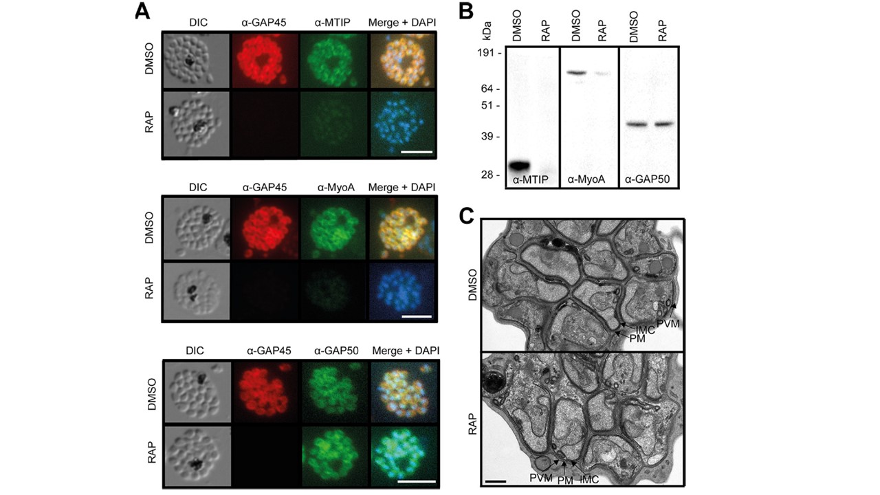
ΔGAP45 parasites show defects in expression of the glideosome components MyoA and MTIP. (A) IFA showing the subcellular localization of GAP45-HA3, MTIP, MyoA, and GAP50 in segmented schizonts of ΔGAP45 (RAP) and mock-treated (DMSO) GAP45:loxP parasites. Loss of GAP45-HA3 also resulted in loss of detection of MTIP and MyoA at the IMC upon RAP treatment. Bars, 5 mm. (B) Western blots showing the decreased overall abundance of MTIP and MyoA proteins in the absence of GAP45. (C) TEM images showing similar merozoite and IMC morphologies in GAP45-HA3 and ΔGAP45 parasites. Merozoite plasma membrane (PM), merozoite IMC, and schizont PVM are indicated. Bar, 0.5 mm. neither MTIP nor MyoA was expressed at wild-type levels in ΔGAP45 schizonts, suggesting that the stable expression of these proteins was dependent upon the presence of GAP45. In contrast, the other major glideosome component, GAP50, was detectable at apparently wild-type levels at the periphery of intracellular ΔGAP45 merozoites. These data demonstrated a defect in motor complex assembly in the ΔGAP45 parasites and support the current model of glideosome assembly.
Perrin AJ, Collins CR, Russell MRG, Collinson LM, Baker DA, Blackman MJ. The Actinomyosin Motor Drives Malaria Parasite Red Blood Cell Invasion but Not Egress. MBio. 2018 Jul 3;9(4). pii: e00905-18.
Other associated proteins
| PFID | Formal Annotation |
|---|---|
| PF3D7_0918000 | glideosome-associated protein 50 secreted acid phosphatase |
| PF3D7_1222700 | glideosome-associated protein 45 |