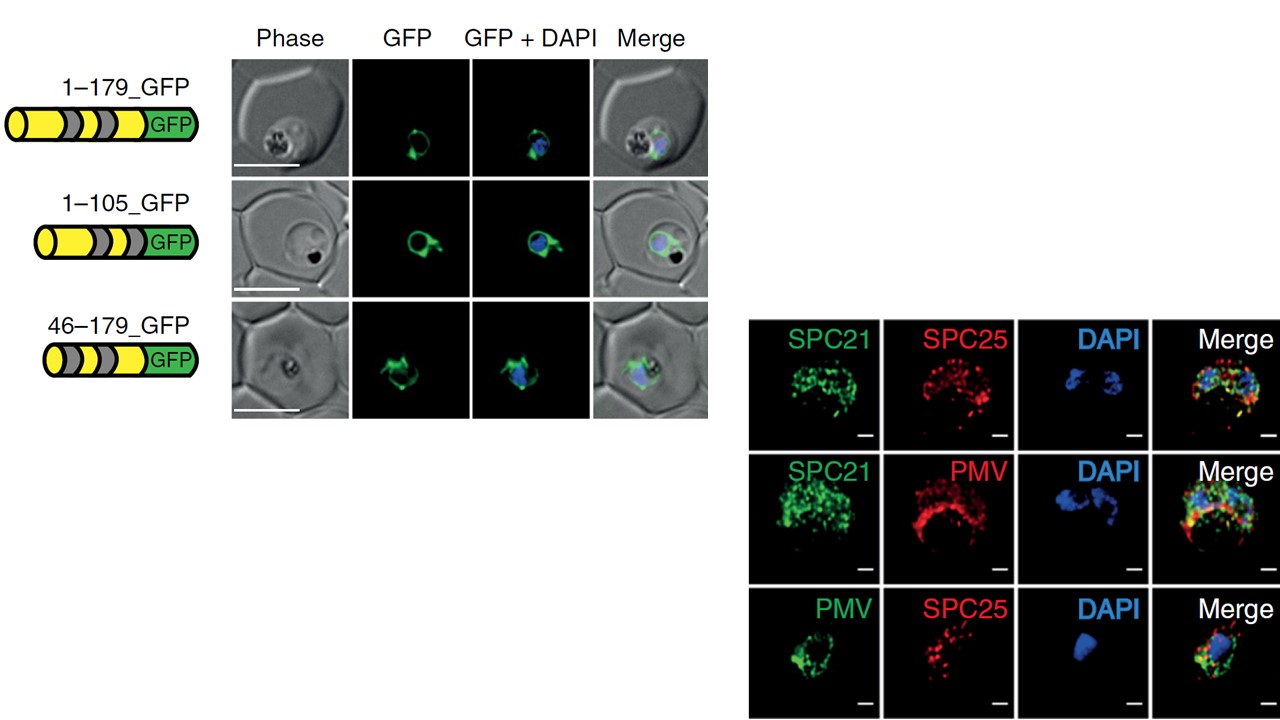
Left: Dissecting the PMV–PfSPC25 interaction. a, A schematic of the SPC25-GFP chimaera (1–179_GFP, 1–105_GFP and 46–179_GFP) and immunofluorescence analysis of all constructs showing that fluorescence localized to the ER. The transmembrane domains are shaded in grey. Nuclei were stained using DAPI. Scale bar, 5 μ m. N = 3 biologically independent samples used. All chimaeras had similar localization to the ER. Right: Characterization of the signal peptidase catalytic subunit PfSPC21. Immunofluorescence analysis of SPC21-HA parasites with anti-HA antibodies. SPC21-HA is colocalized at the ER with anti-SPC25 (top row) and anti-PMV (bottom row). Nuclei were localized with DAPI. Scale bars, 1 μ m. More than ten parasites were imaged for each condition. N = 3 biologically independent samples used.
Marapana DS, Dagley LF, Sandow JJ, Nebl T, Triglia T, Pasternak M, Dickerman BK, Crabb BS, Gilson PR, Webb AI, Boddey JA, Cowman AF. Plasmepsin V cleaves malaria effector proteins in a distinct endoplasmic reticulum translocation interactome for export to the erythrocyte. Nat Microbiol. 2018 Aug 20. [Epub ahead of print]
Other associated proteins
| PFID | Formal Annotation |
|---|---|
| PF3D7_1323500 | PEXEL protease plasmepsin V |
| PF3D7_1345900 | kinetochore protein SPC25, putative |