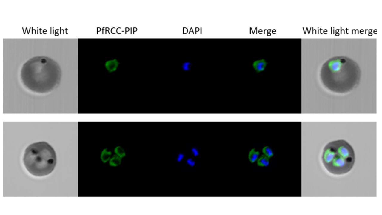
Cellular distribution of PfRCC-PIP-HA analyzed by immunofluorescence with anti-HA antibodies. The PfRCC-PIP protein appears in green and the nucleus is stained in blue by DAPI. Upper and lower panels show one erythrocyte infected by one trophozoite and 3 trophozoites, respectively. The fluorescent signal is detected at the periphery of the nucleus.
Lenne A, De Witte C, Tellier G, Hollin T, Aliouat EM, Martoriati A, Cailliau K, Saliou JM, Khalife J, Pierrot C. Characterization of a Protein Phosphatase Type-1 and a Kinase Anchoring Protein in Plasmodium falciparum. Front Microbiol. 2018 9:2617