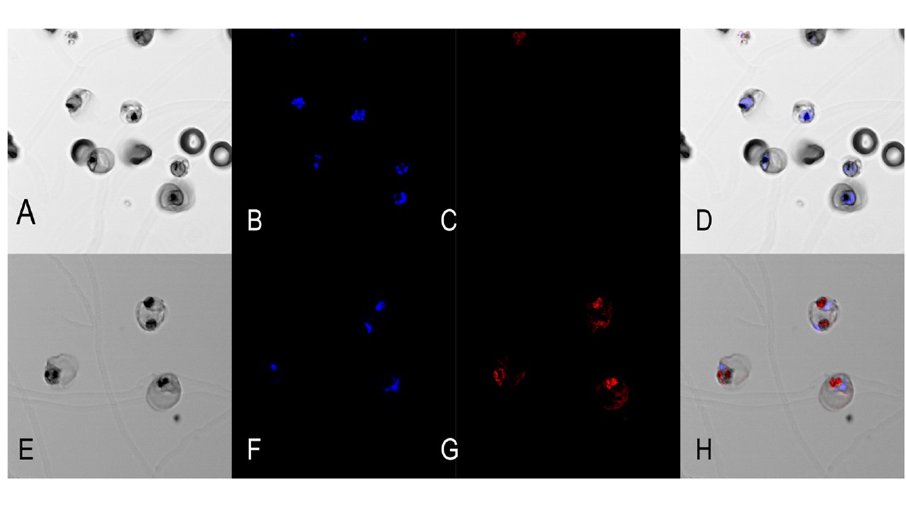
Immunofluorescence assay using anti-PFI1140w and anti-PF13_0353 antisera. Purified P. falciparum trophozoites were incubated with a 1/500 dilution of antisera
recognizing coding sequences of either PFI1140w or PF13_0353. Goat anti-mouse IgG conjugated with Alexa 633 was used as a secondary antibody. Nuclear DNA was labeled with Hoescht 33258. a: DIC image of trophozoites treated with anti-PFI1140w antiserum b: nuclear DNA stained with Hoescht 33258 and c: negative Alexa 633 signal. d: merged images of a, b and c. e: DIC image of trophozoites treated with anti-PF13_0353 antiserum, f: nuclear DNA stained with Hoescht 33258 and g: Alexa 633 signal recognizing anti- PF13_0353 antibody. h: merged images of e, f and g. Immunofluorescence microscopy assays (IFA) with antiserum against PFI1140w failed to show reactivity with parasitic structures (Fig 9 a, b, c and d). On the contrary, when anti-PF13_0353 antiserum was tested by IFA in trophozoites, it showed positive fluorescence signals that did not overlap with nuclei detected by Hoescht 33258 (Fig 9 e, f, g and h). In addition, the observed reactivity of anti-PF13_0353 antiserum was localized in the membranous region surrounding the food vacuole area.
Ostera G, Tokumasu F, Oliveira F, Sa J, Furuya T, Teixeira C, Dvorak J. Plasmodium falciparum: food vacuole localization of nitric oxide-derived species in intraerythrocytic stages of the malaria parasite. Exp Parasitol. 2008 120(1):29-38.
Other associated proteins
| PFID | Formal Annotation |
|---|---|
| PF3D7_1367500 | NADH-cytochrome b5 reductase, putative |