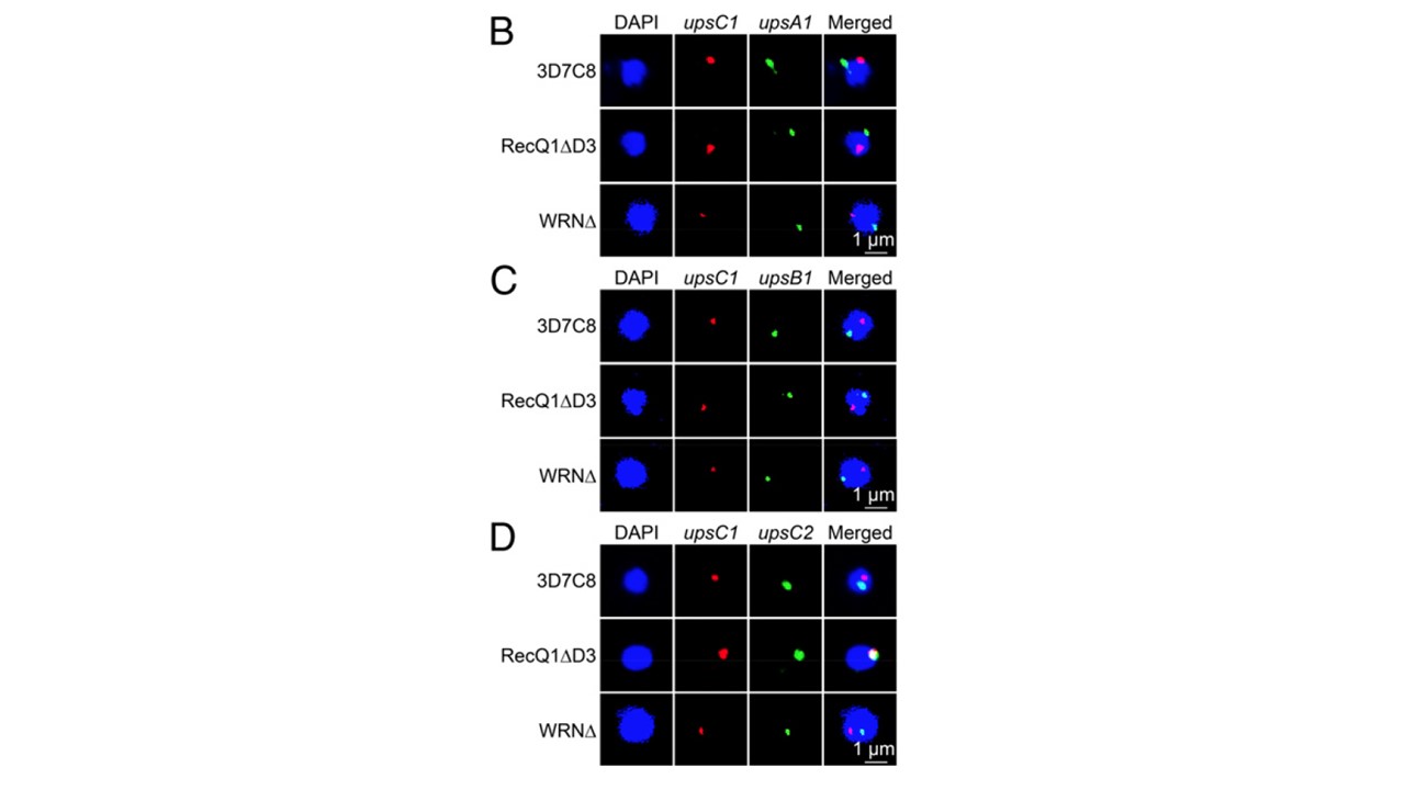
Representative image of paired DNA-FISH of upsA1 (PF3D7_0400400, green) with upsC1 (PF3D7_1240600, red). In 3D7C8 and RecQ1Δ, upsA1 was labeled with fluorescein, whereas upsC1 was labeled with biotin. In WRNΔ, upsA1 was labeled with biotin, whereas upsC1 was
labeled with fluorescein. (C) Representative image of paired DNA-FISH of upsB1 (PF3D7_ 0115700, green) with upsC1 (red). In all strains, upsB1 was labeled with biotin, whereas upsC1 was labeled with fluorescein. (D) Representative image of paired DNA-FISH of upsC2 (PF3D7_0412400, green) with upsC1 (red). In 3D7C8 and RecQ1Δ, upsC2 was labeled with fluorescein and upsC1 was labeled with biotin. In WRNΔ, upsC2 was labeled with biotin
and upsC1 was labeled with fluorescein. Parasites synchronized at ring stage were used for this DNA-FISH. Biotin labeling was visualized by incubating with streptavidin-Alexa Fluor 568. DAPI staining (blue) was used to locate nuclear area. upsC1 was predominantly not colocalized with those of upsA1 or upsB1 in both WT 3D7 and PfRecQ1Δ, suggesting the spatial
distribution of upsC1 relative to the upsA or upsB type was unaffected by deletion of PfRecQ1 (B and C). Interestingly, upsC1 became predominantly colocalized with upsC2 in PfRecQ1Δ (4).
Li Z, Yin S, Sun M, Cheng X, Wei J, Gilbert N, Miao J, Cui L, Huang Z, Dai X, Jiang L. DNA helicase RecQ1 regulates mutually exclusive expression of virulence genes in via heterochromatin alteration. Proc Natl Acad SciPlasmodium falciparum U S A. 2019 116(8):3177-3182.
Other associated proteins
| PFID | Formal Annotation |
|---|---|
| PF3D7_0400400 | erythrocyte membrane protein 1, PfEMP1 |
| PF3D7_0918600 | ATP-dependent DNA helicase Q1; bloom protein homolog |
| PF3D7_1240600 | erythrocyte membrane protein 1, PfEMP1 |