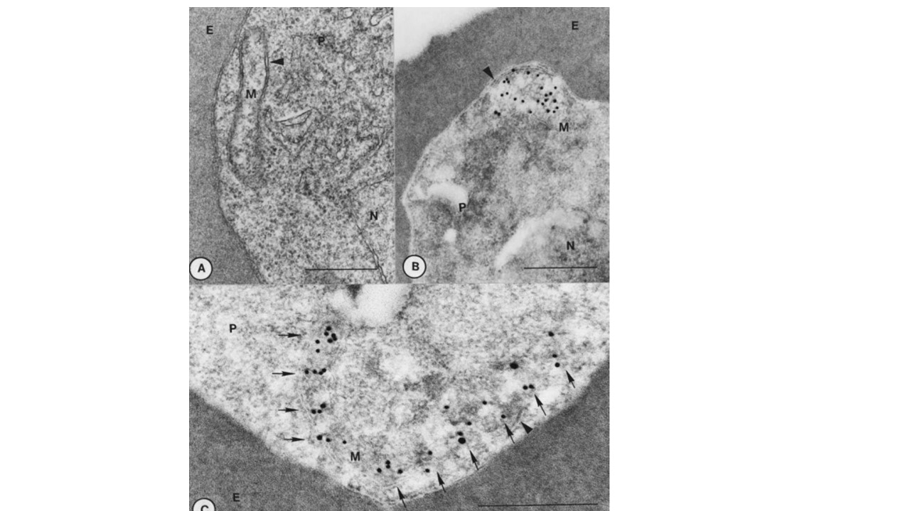
Immunoelectron microscopic localization of PfHsp60; (A) Epon-embedded section of a P. falciparum trophozoite (P) showing an acristate mitochondrion (M). It is composed of outer and inner membranes (arrow head). E, erythrocyte; N, nucleus.Bar=0.5 mm; (B) LR white section of a P. falciparum trophozoite (P). A mitochondrion (M) is associated with gold particles indicating the localization of PfHsp60 molecules. Mitochondrion is composed of double membranes (arrow head). E, erythrocyte; N, nucleus. Bar=0.5 mm; (C) LR white section showing a mitochondrion (M) of P. falciparum (P) infected erythrocyte (E). Mitochondrion is composed of double membranes (arrow head). Gold particles showing band pattern (arrows) may indicate compartmental localization of PfHsp60. Bar=0.5 mm.
Das A, Syin C, Fujioka H, Zheng H, Goldman N, Aikawa M, Kumar N. Molecular characterization and ultrastructural localization of Plasmodium falciparum Hsp 60. Mol Biochem Parasitol. 1997 88(1-2):95-104.