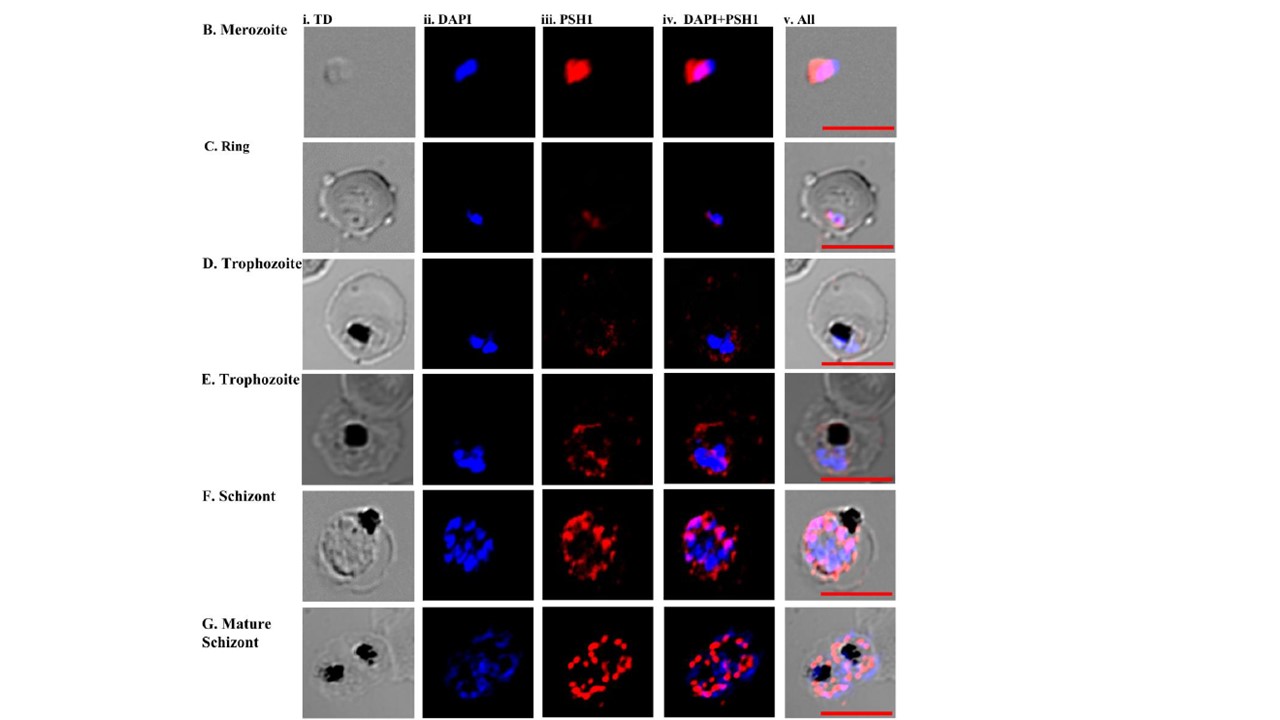
Localization of PfPSH1 in different intra-erythrocytic stages of P. falciparum. (A) Staining with pre-immune sera (i) phase contrast (TD) image; (ii) image of cell stained with DAPI (blue); (iii) pre-immune sera; (iv) DAPI + pre-immune sera (v) All merged;(B-F) Staining with anti-PSH1 sera (B) merozoite stage, (C) ring stage, (D and E) trophozoite stage, (F) schizont stage, (G) mature schizont stage (i) phase contrast (TD) image; (ii) DAPI stained cells (blue); (iii) immunofluorescent stained cell Alexa 594 (PSH1); (iv) Merged image of panel ii and iii; (v) Merged image of panel i-iv; In A-G red scale bar indicates the size 5 micro meters. anti-PfPSH1N antibodies show its localization in both nucleus and cytoplasm. In early stages PfPSH1 is located in both cytoplasm and nucleus such as in merozoite it is present in cytoplasm and partially in nucleus (panels i-v) and incase of ring stage it is present in cytoplasm and very less in nucleus (Fig. 6C, panels i-v). In trophozoite stage it is majorly present in the cytoplasm (Fig. 6D and 6E, panels i-v). In later developmental stages like schizont stage it is mostly located in nucleus as compared to cytoplasm (F and G, panels i-v).
Chauhan M, Sourabh S, Yasmin R, Pahuja I, Tuteja R. Biochemical characterisation of Plasmodium falciparum parasite specific helicase 1 (PfPSH1). FEBS Open Bio. 2019 Aug 30