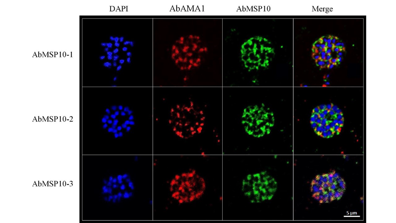
Evaluation of AbMSP10 by confocal microscopy. Purified schizont smears were fixed onto microscopy slides and incubated with antibodies AbMSP10-1, AbMSP10-2 or AbMSP10-3 at a concentration of 1 μg/mL. A strong fluorescence signal was observed for antibodies AbMSP10-1 and AbMSP10-2. AbMSP10-3 showed a dot pattern noisy signal probably due to the reduced specificity of this antibody. AbMSP10 (green), AbAMA1 (red), the nucleus of merozoites are blue (DAPI).
Bendezu J, Villasis E, Morales Ruiz S, Garro K, Infante B, Gutierrez-Loli R, Rodríguez P, Fernández-Díaz M, Gamboa D, Torres K. Evaluation of Plasmodium falciparum MSP10 and its development as a serological tool for the Peruvian Amazon region. Malar J. 2019 18(1):327.
PubMed Article: Evaluation of Plasmodium falciparum MSP10 and its development as a serological tool for the Peruvian Amazon region
Other associated proteins
| PFID | Formal Annotation |
|---|---|
| PF3D7_1133400 | apical membrane antigen 1 |