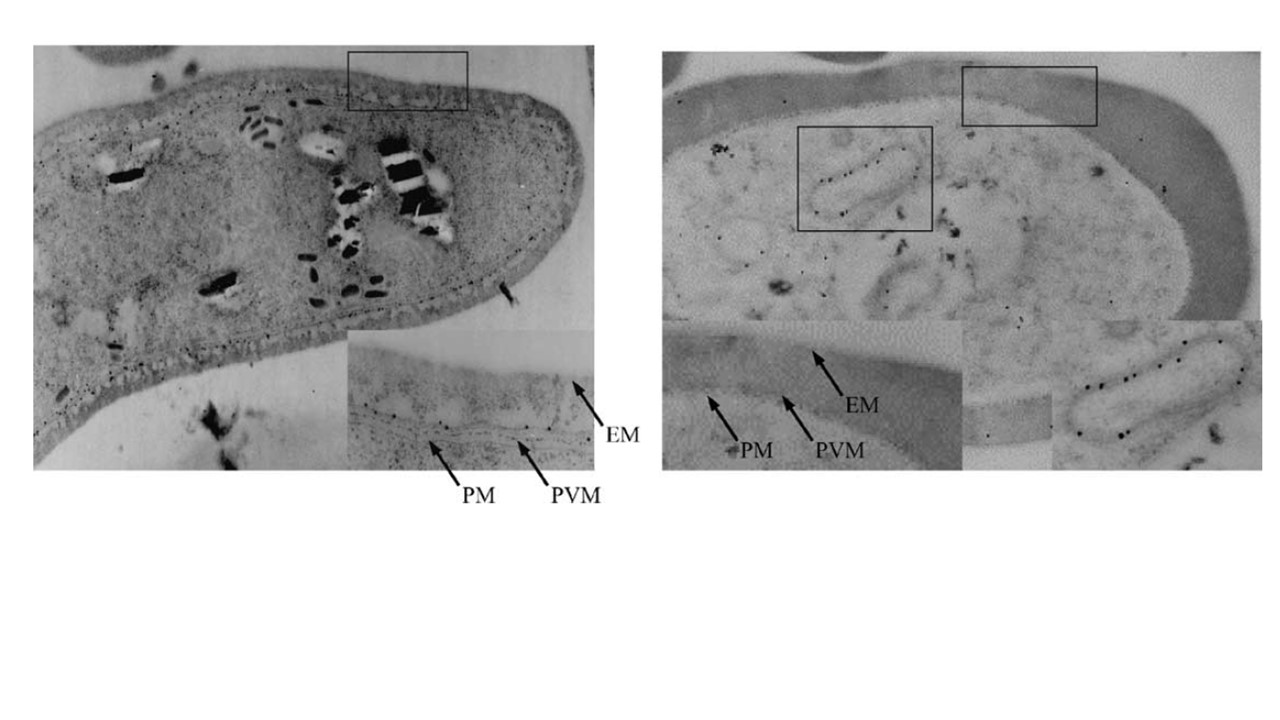
Immuno-electron microscopy on gametocytes prepared from wild type parasites (left panel) and from 16ko-2 parasites (right panel). Various membranes are marked as EM (erythrocyte membrane), PVM (parasitophorous vacuole membrane), and PM (plasma membrane). Inset in (C) shows a higher magnification of PVM localization of Pfs16 in wt 3D7. Inset in (D) shows higher magnification of altered localization of proteins recognized by Pfs16 specific antibodies in the 16ko parasites and the lack of Pfs16 localization on the PVM. WT parasites showed localization of Pfs16 to be distributed throughout the PVM. “16ko” parasites clearly showed that the putative truncated Pfs16 protein did not traffic to the PVM, instead the putative truncated protein was retained inside the cytoplasm in an unidentified subcellular compartments with double membrane layers.
Kongkasuriyachai D, Fujioka H, Kumar N. Functional analysis of Plasmodium falciparum parasitophorous vacuole membrane protein (Pfs16) during gametocytogenesis and gametogenesis by targeted gene disruption. Mol Biochem Parasitol. 2004 133(2):275-85