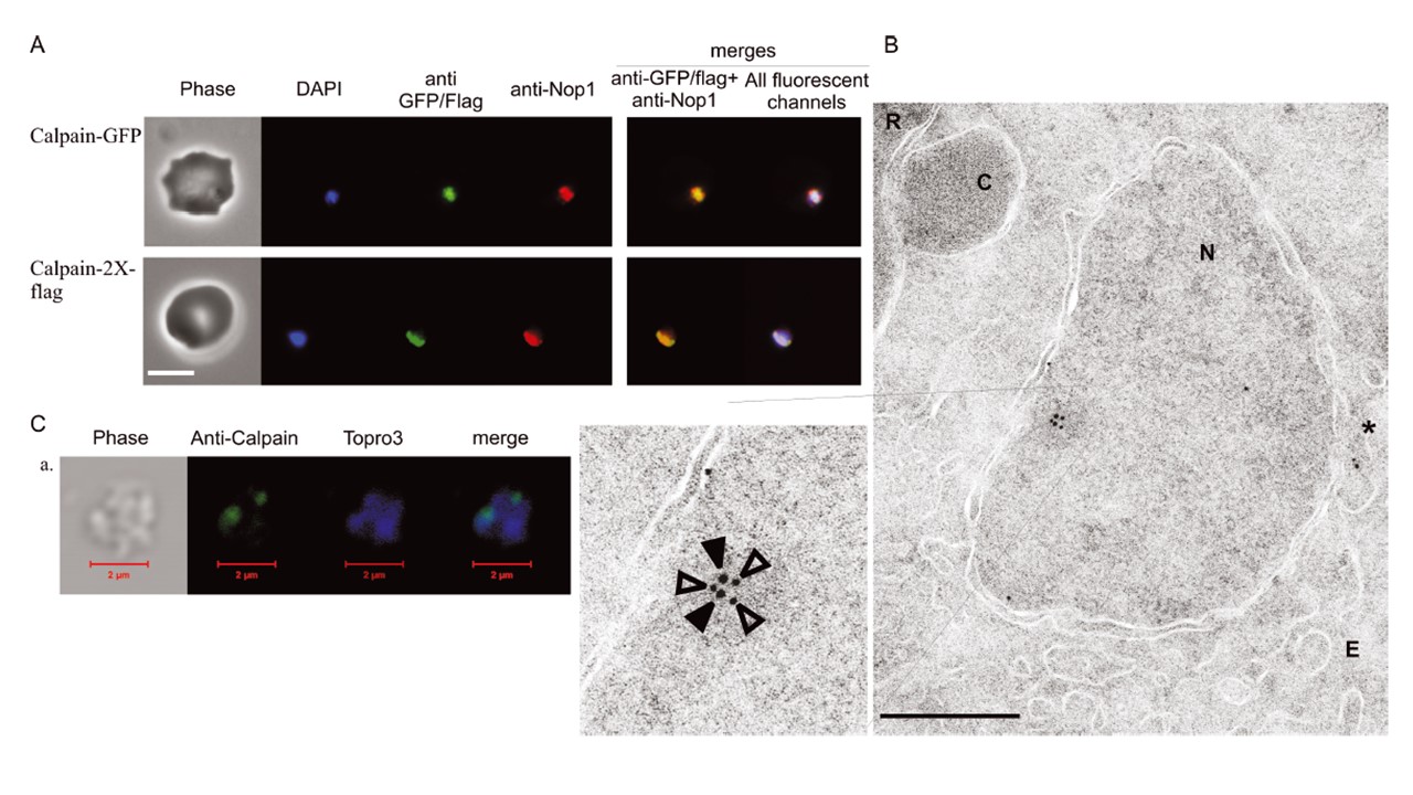
Immunofluorescence and EM analyses reveal that Pf calpain is nucleolar. A. Colocalization of calpain and Nop1, a nucleolar marker. Using specific antibodies for GFP or for flag epitope in combination with proper AF488-conjugated secondary antibodies, we detected the cellular distribution of calpain in the clones expressing calpain-GFP and calpain-2X-flag in relation to the nucleolar compartment labelling obtained with an anti-Pf_Nop1 serum (red channel). The nuclei are stained with DAPI. Bar, 5 mm. B. Immuno-EM was done using pre-embedding labelling with anti-flag and anti-Pf_Nop1 after tetanolysin treatment to permeabilize erythrocytes. The image presents the nuclear section of a representative early trophozoite. At the upper left corner are visible the red blood cell (R) and a cytostome (C). The nucleus (N) appears thoroughly circumscribed by its double membrane closely associated with ER (E). Inside the nucleus, labelling due to the immunodetection of calpain-flag (gold particles 12 nm – open arrows in the enlarged panel) and Pf_Nop1 (gold particles 18 nm – solid arrows in the enlarged panel) reveals staining of an electron-dense nuclear subcompartment. Some particles are also found in the perinuclear ER (asterisk). Bars, 0.5 mm. C. Confocal analysis of purified nuclei from 3D7 using anti-calpain antiserum #35. We generally detected one or two spots of approximate diameter of 0.5 mm. Colocalizazion of Nop1 and calpain in an isolated nucleus by confocal analysis is provided in Fig. S3.
Russo I, Oksman A, Goldberg DE. Fatty acid acylation regulates trafficking of the unusual Plasmodium falciparum calpain to the nucleolus. Mol Microbiol. 2009 72(1):229-45
Other associated proteins
| PFID | Formal Annotation |
|---|---|
| PF3D7_1407100 | rRNA 2'-O-methyltransferase fibrillarin, putative |