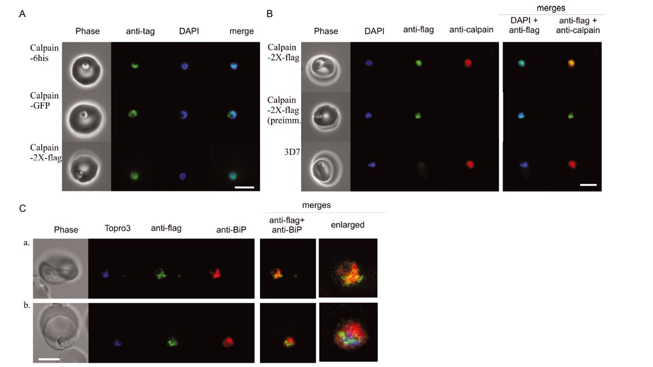
Immunofluorescence localization of calpain. A. Immunofluorescence images of clones expressing calpain-6-his, calpain-GFP and calpain-2X-flag are presented. The images are representative of the overall population and were obtained using tag-specific primary antibodies and a proper secondary antibody conjugated to Alexa-Fluor (AF) 488. The immunofluorescence distribution in all the clones highlights a region closely associated with the nucleus but not completely merging with the DAPI signal. B. Colocalization of fluorescence signal distributions from both anti-flag antibody and anti-calpain antibody with that of a C-terminal peptide of calpain. Images a and b show the calpain-2X-flag clone stained with monoclonal anti-flag antibody/goat AF488-conjugated anti-mouse antibody and either rabbit anti-calpain #35/goat AF555-conjugated anti-rabbit or pre-immune serum of the same rabbit/goat AF555-conjugated anti-rabbit. The 3D7 parental strain, stained as in a, is positive with anti-calpain #35, but not with the anti-flag antibody. C. The relative distributions of calpain-2X-flag (green channel) and BiP (red channel) signals were analysed by confocal microscopy. Here two representative images show the signal detected in a perinuclear plane (a) and in a nuclear plane (b). The right-most panels present the enlarged merge images. The nucleus in (a) was detected acquiring the Topro3-signal with increased pin-hole that rendered a non-confocal image. Bars, 5 mm.
Russo I, Oksman A, Goldberg DE. Fatty acid acylation regulates trafficking of the unusual Plasmodium falciparum calpain to the nucleolus. Mol Microbiol. 2009 72(1):229-45.
Other associated proteins
| PFID | Formal Annotation |
|---|---|
| PF3D7_1362400 | calpain |