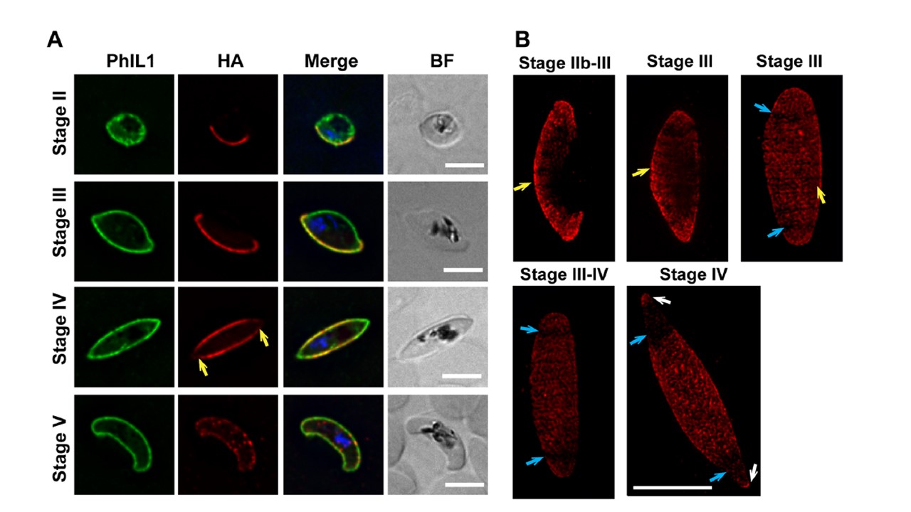
PhIL1-interacting protein, PIP1, is located at the gametocyte IMC and is essential for gametocyte development. (A) PIP1-HA-glmS transfectants were labeled with anti-PhIL1 rabbit (green) and anti-HA (red) at different developmental stages (II±V). Nuclei were labeled with DAPI. There is an apparent reduction in fluorescence intensity of PIP1-HA labeling at the tips of the gametocyte in stage IV (yellow arrow). In stage V, the fluorescence becomes punctate at the parasite periphery, while PhIL1 remains homogenous. (B) 3D-SIM imaging of PIP1-HA-glmS during gametocyte development. Homogeneous labeling of the IMC plates by anti-HA (red) are observed (yellow arrows) in the early stages of development. During stage III±IV of development, there is a loss of labeling from the plates near the tips of the elongated parasite (blue arrows), with some protein labeling of the tips themselves (white arrows). Scale bar: 5 μm.
Parkyn Schneider M, Liu B, Glock P, Suttie A, McHugh E, Andrew D, Batinovic S, Williamson N, Hanssen E, McMillan P, Hliscs M, Tilley L, Dixon MWA. Disrupting assembly of the inner membrane complex blocks Plasmodium falciparum sexual stage development. PLoS Pathog. 2017 13(10):e1006659.
Other associated proteins
| PFID | Formal Annotation |
|---|---|
| PF3D7_0109000 | photosensitized INA-labeled protein PHIL1 |