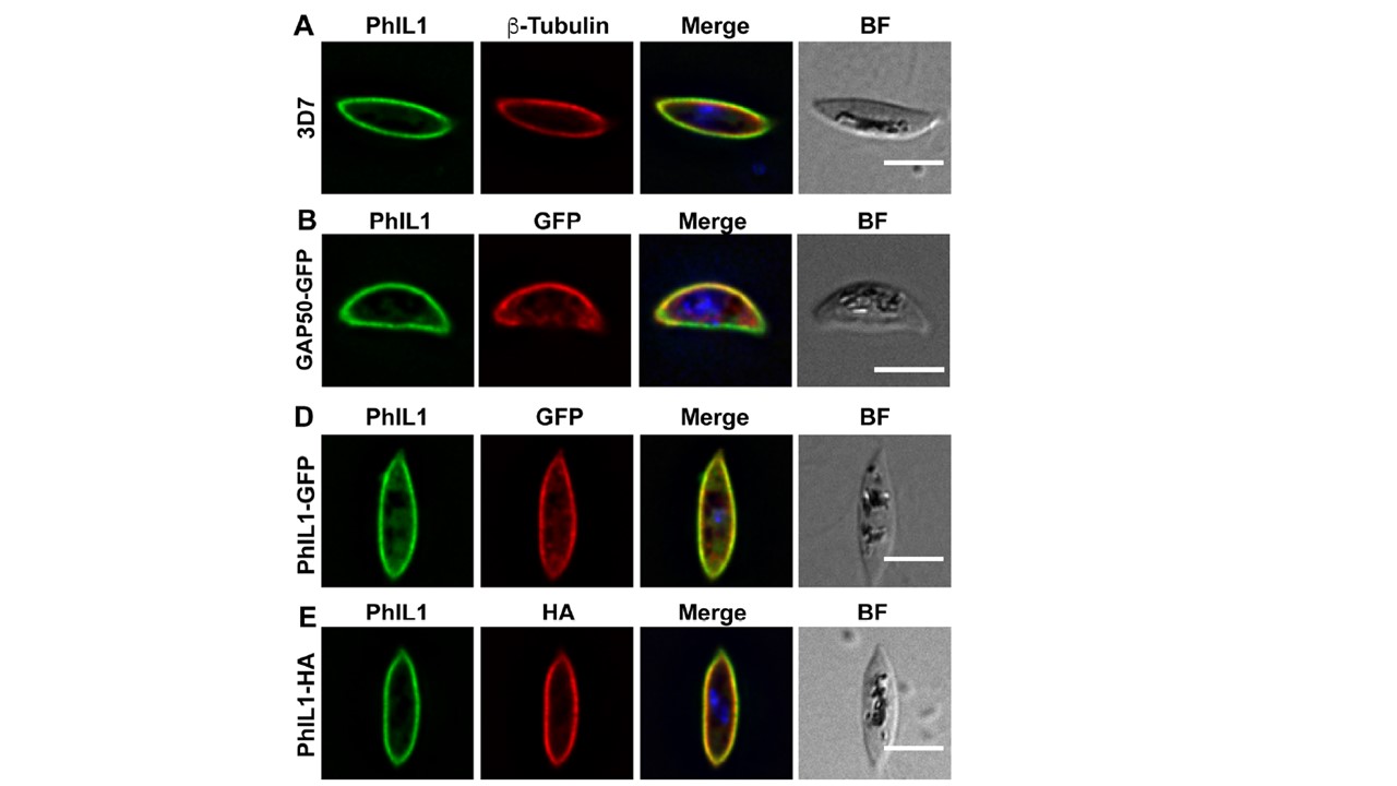
PhIL1 is located at the IMC in gametocytes. (A) Immunofluorescence microscopy showing anti-PhIL1 (green) at the periphery of a 3D7 stage IV gametocyte, showing fluorescence close to the anti-β-tubulin (red) labeling. Immunofluorescence microscopy of stage IV gametocytes revealed a strong PhIL1 signal at the periphery, where it is closely associated with β-tubulin and was located close to the known IMC marker, the glideosome-associated protein 50 (B) Immunofluorescence microscopy of a stage IV gametocyte showing overlap of GAP50-GFP (red) and PhIL1 (green) at the periphery of the cell. (D, E) Immunofluorescence microscopy of stage IV PhIL1-GFP (D) and PhIL1-HA (E) gametocyte transfectants. Parasites were labeled with anti-PhIL1 (green) and anti-GFP or anti-HA (red). Nuclei were labeled with DAPI. Scale bars: 5 μm. A PhIL1-HA line was also generated by integrating 3xHA into the 3' end of the PhIL1 locus. Immunofluorescence signals for HA and GFP overlapped with the signal from anti-PhIL1 antibody, confirming the correct location of the tagged proteins (D and E).
Parkyn Schneider M, Liu B, Glock P, Suttie A, McHugh E, Andrew D, Batinovic S, Williamson N, Hanssen E, McMillan P, Hliscs M, Tilley L, Dixon MWA. Disrupting assembly of the inner membrane complex blocks Plasmodium falciparum sexual stage development. PLoS Pathog. 2017 13(10):e1006659.
Other associated proteins
| PFID | Formal Annotation |
|---|---|
| PF3D7_0918000 | glideosome-associated protein 50 secreted acid phosphatase |
| PF3D7_1008700 | tubulin beta chain |