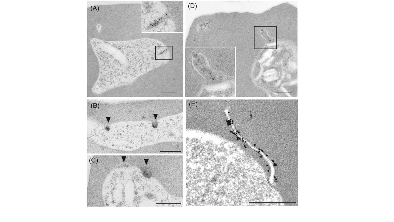
Subcellular localization of SBP1 by immunoelectron microscopy using sections of early phase P. falciparum erythrocytic stage development parasites. (A) Gold particles indicating the localization of SBP1 are observed in the electron dense material (EDM) in the cytoplasm of an early trophozoite. Inset: larger magnification image of EDM area. Bar, 500 nm. (B) Two EDMs where SBP1 is localized are observed in contact with PPM (Arrowhead). Bar, 500 nm. (C) Gold particles showing
deposition of SBP1 are observed in EDM located between PPM and PVM (Arrowhead). Bar, 500 nm. (D) PVM in contact with the EDM is invaginating toward the cytoplasmic side of the trophozoite infected erythrocyte. Inset: larger magnification image of EDM. Bar, 500 nm. (E) Gold particles are deposited on a newly formed Maurer's cleft located adjacent to PVM. Bar, 500 nm. SBP1 is inserted into the Maurer's cleft with the N-terminus facing the lumen side of the cleft and the C-terminus facing the erythrocyte cytoplasm. In this study IEM showed that gold particles conjugated with antibodies against the C-terminus of SBP1 accumulate inside the electron dense material (EDM) of the PV space (D) and outside the Maurer's clefts near the PVM (E). These findings suggest that SBP1 initially accumulates in EDM and then is arranged with the C-terminus exposed to the erythrocyte cytoplasm during Maurer's cleft formation.
Iriko H, Ishino T, Tachibana M, Omoda A, Torii M, Tsuboi T. Skeleton binding protein 1 (SBP1) of Plasmodium falciparum accumulates in electron-dense material before passing through the parasitophorous vacuole membrane. Parasitol Int. 2019 Nov 13;75:102003.