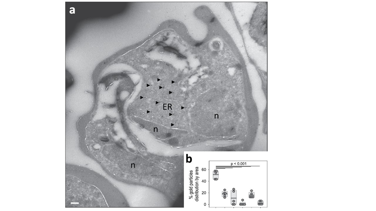
Subcellular localization of PfUT by immunoelectron microscopy. (a) A representative micrograph is shown of an erythrocyte infected with a pfut mutant. The sample was prepared according to the Tokuyasu protocol, and immunolabelled with anti-HA antiserum (mouse, 1:1), followed by staining with an anti-mouse antibody coupled to 10 nm colloidal gold (goat, 1:20). Arrowheads point towards gold label. n, nucleus; ER, endoplasmic reticulum/Golgi complex. Scale bar, 200 nm. (b) Quantification of immuno EM results. The distribution of gold grains was determined in 5 micrographs and analyzed according to their subcellular localization. Gold grains were significantly more present in areas of the ER/Golgi complex (ER) than in other subcellular compartments, including the parasite’s cytoplasm (C), nucleus (N), digestive vacuole (DV), red blood cell cytosol (RBC) and non-cellular background (BG). Each symbol represents data derived from an individual parasitized erythrocyte. A box plot analysis is overlaid over the individual data points, with the median (thin grey line), mean (thick black line) and the 25% and 75% quartile ranges being shown. Statistical significance between the data sets was assessed using the Holm-Sidak one way ANOVA test. Immuno-electron microscopy using anti-HA antiserum confirmed a subcellular localization of PfUT at the ER/Golgi complex in the mutant lines. A comparison of the micrographs with reference electron microscopic images of trophozoites provided no evidence of morphological abnormalities in the mutant parasite lines, suggesting normal phenotypic characteristics of organelles and other subcellular compartments.
Jankowska-Döllken M, Sanchez CP, Cyrklaff M, Lanzer M. Overexpression of the HECT ubiquitin ligase PfUT prolongs the intraerythrocytic cycle and reduces invasion efficiency of Plasmodium falciparum. Sci Rep. 2019 Dec 4;9(1):18333.