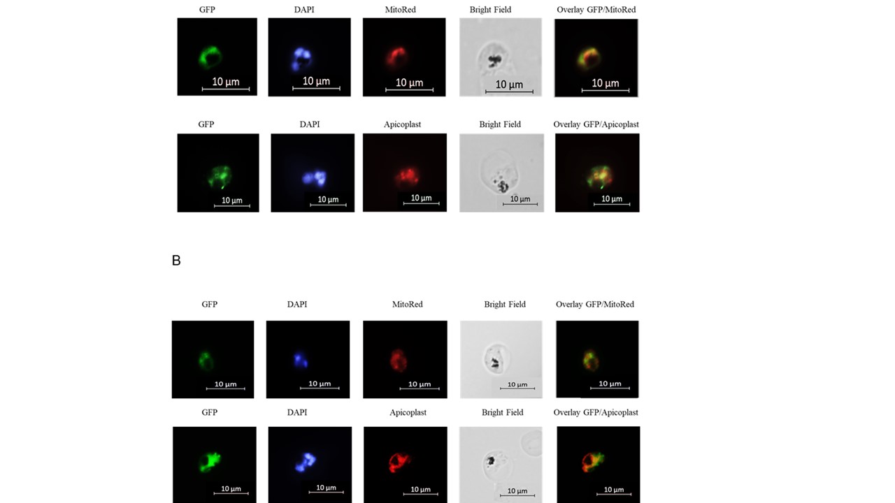
The localization of GFP-tagged COX10. Parasites labeled with DAPI and MitoRed (upper panels A and B) or with an apicoplast-specific antibody (bottom panels, A and B) formulated according to Tonkin et al.,30. A and B represent exposures of different parasites. The COX10 images show a larger area of GFP overlap with the mitochondria (MitoRed, upper panels A and B), a small overlapping area with the nucleus (DAPI) and an overlap with the apicoplast that exceeds this organelle’s limits, indicating that the enzyme is in a larger organelle (anti-apicoplast, bottom panels A and B). Note that in dividing cells, Acyl carrier protein (ACP)-carrying apicoplasts are seen as distinct spots which are not directly coincident with the COX10 signal (green), suggesting a differential localization of PfCOX10 and ACP in apicoplasts. Pictures were taken with a Zeiss LSM-780 NLO Multifoot confocal.
Simão-Gurge RM, Wunderlich G, Cricco JA, Cubillos EFG, Doménech-Carbó A, Cebrián-Torrejón G, Almeida FG, Cirulli BA, Katzin AM. Biosynthesis of heme O in intraerythrocytic stages of Plasmodium falciparum and potential inhibitors of this pathway. Sci Rep. 2019 Dec 17;9(1):19261
Other associated proteins
| PFID | Formal Annotation |
|---|---|
| PF3D7_0519300 | protoheme ix farnesyltransferase COX10 |