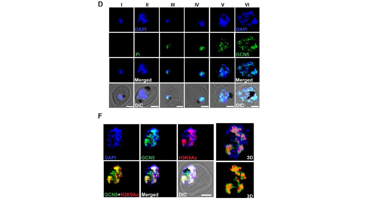
Expression analysis of PfGCN5 in asexual blood stages. (D) Immunofluorescence analysis of PfGCN5 in different parasite stages using pre-immune sera (I and II) and immune PfGCN5sera (III, IV, V, and VI). Alexa fluor 488–tagged secondary antibodies were used for the detection of PfGCN5. DAPI was used to stain the nucleus. Pre-immune sera did not show any signal from the parasite [D (I & II)]. Figure D shows the expression of PfGCN5 in all the stages. During early stages of development PfGCN5 signal was distributed over the nuclear DAPI signal [D (III & IV)] that showed more punctate pattern over the nucleus during later stages of development [D (V & VI)]. (F) Co-localization of PfGCN5 and PfH3K9Ac in fixed parasites using antibodies against respective proteins. The nuclei were stained with DAPI. Alexa fluor 488 and Alexa fluor 594-tagged secondary antibodies were used to detect PfGCN5 (green) and PfH3K9Ac (red) respectively. 3D view of selected images was shown on extreme right for better clarity. The results indicated a significant co-localization between these two proteins (Pearson's correlation coefficient is 0.79). Some independent PfGCN5 signal were also observed that may reflect separate function of PfGCN5 apart from its histone acetylation function.
Bhowmick K, Tehlan A, Verma S, Sudhakar R, Kaur I, Sijwali PS, Krishnamachari A, Dhar SK. Plasmodium falciparum GCN5 acetyltransferase follows a novel proteolytic processing pathway essential for its function. J Cell Sci. 2019 Dec 20. pii: jcs.236489.