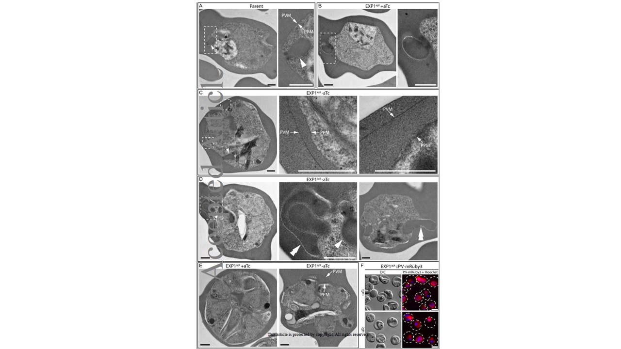
Depletion of EXP1 results in PV/PVM morphological abnormalities. (A-E) TEM visualization of parasite ultrastructure in parent and EXP1apt parasites grown 48 hours with or without aTc. Images are shown of (A-D) trophozoites and (E) segmented schizonts. Dashed boxes indicate enlarged areas shown to the right. Arrows indicate PVM and PPM. Arrowheads indicate cytostomes. Double arrowheads indicate abnormal membrane-enclosed structures filled with host cytosol in the PV lumen. Results are representative of two independent experiments. Scale bars are 500 nm. (F) Live fluorescence imaging of magnet-purified EXP1apt::PV-mRuby3 parasites grown 48 hours with or without aTc. The dashed lines indicate the boundary of infected RBC traced from the corresponding DIC image. Results are representative of three independent experiments. Scale bars are 5 μm.
Nessel T, Beck JM, Rayatpisheh S, Jami-Alahmadi Y, Wohlschlegel JA, Goldberg DE, Beck JR. EXP1 is required for organization of EXP2 in the intraerythrocytic malaria parasite vacuole. Cell Microbiol. 2020 Jan 28.