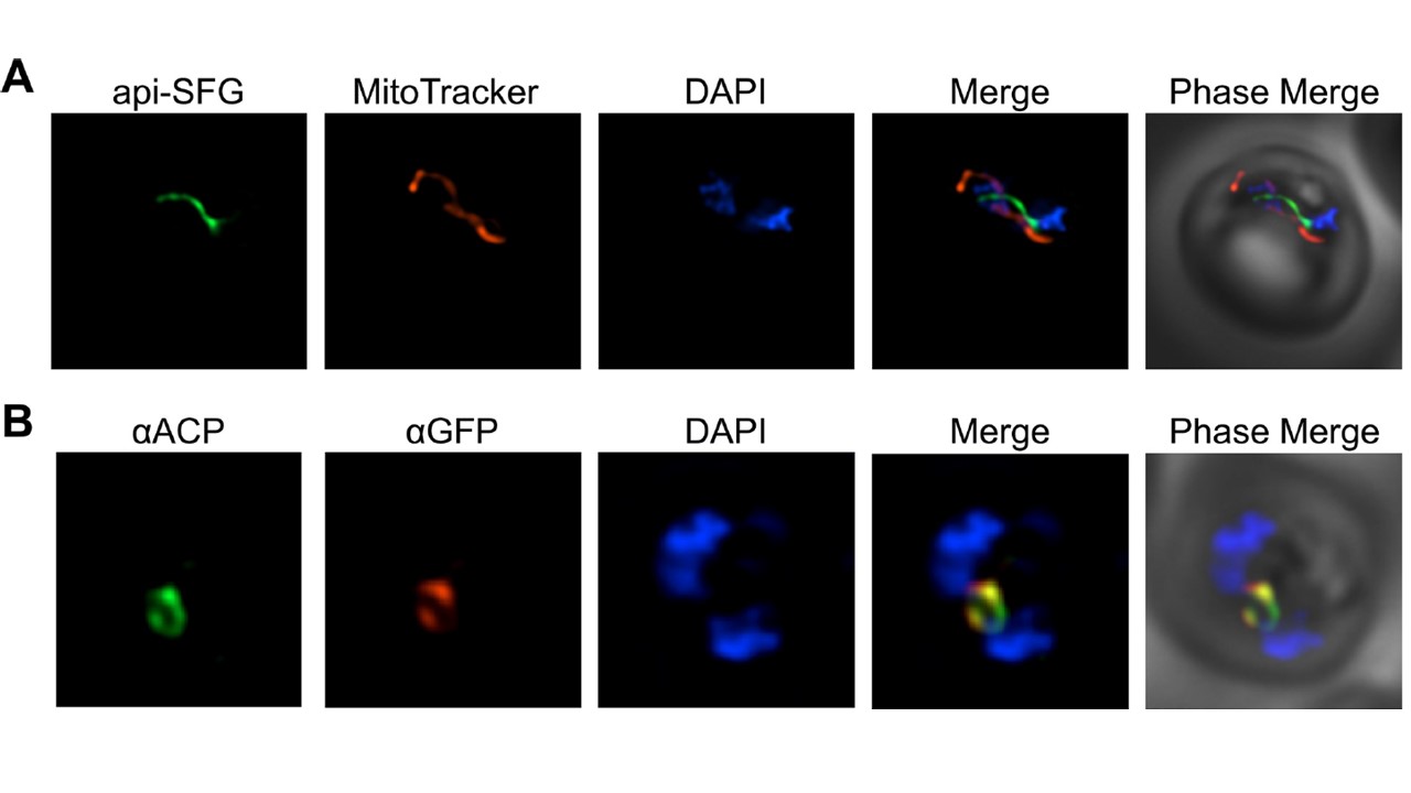
Fluorescent labeling of the apicoplast organelle in PfMev parasites and confirmation via co-localization. A) Live fluorescence microscopy of the PfMev line (express the mevalonate pathway enzymes expressing the signal sequence and transit peptide from ACP fused to SFG (green: Super Folder Green), the mitochondria was stained with MitoTracker (red), and nuclear DNA was stained with DAPI (blue). B) Immunofluorescence co-localization of aSFG with the apicoplast marker ACP, with α-ACP (green), α-GFP (red), with nuclear DNA stained with DAPI (blue) in fixed PfMev parasites. Images depict fields that are 10 microns long by 10 microns wide. SFG is trafficked to a subcellular compartment consistent with the morphology of the apicoplast organelle (A). Trafficking of api-SFG to the apicoplast was confirmed using immunofluorescence (IFA) by co-localizing api-SFG with ACP–a known marker of the apicolast organelle (B).
Swift RP, Rajaram K, Liu HB, et al. A mevalonate bypass system facilitates elucidation of plastid biology in malaria parasites. PLoS Pathog. 2020;16(2):e1008316.