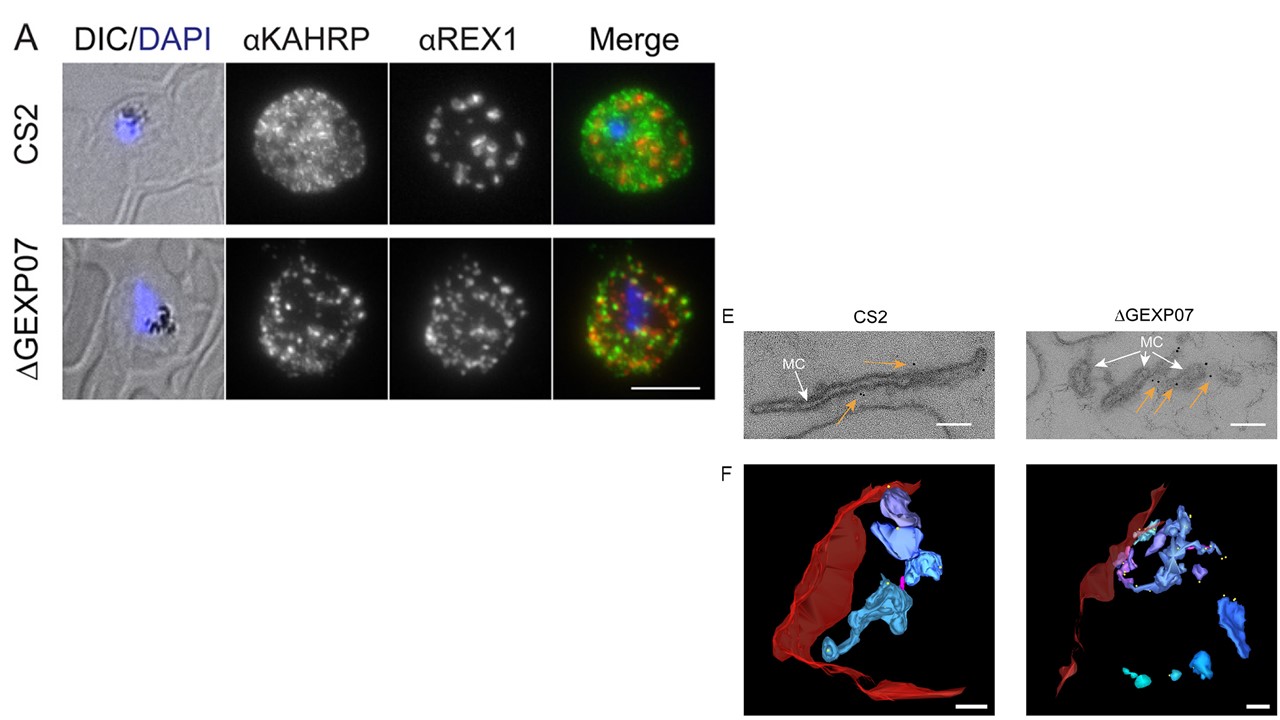
Knockout of GEXP07 alters parasite growth and Maurer’s cleft architecture. (A) Infected RBCs were fixed and probed with anti-KAHRP (green) and counter stained with anti-REX1 (red). Nuclei were stained with DAPI (blue). CS2=parent line. Projections of Z stacks are shown. Scale bar=5 mm. (E) EqtII-permeabilized wild-type CS2 and GEXP07 knockout transfectants were labeled with antibodies recognizing REX1 followed by immunogold labeling and were prepared for electron microscopy. Images have been cropped around Maurer’s clefts (white arrows). Additional images displaying the Maurer’s cleft morphologies are shown in Fig. S6A. Gold arrows point to gold particles. Scale bar 100 nm. (F) Rendered 3D models of Maurer’s clefts generated from electron tomograms. Red = RBC; magenta stalk = tether; pastel hues = independent clefts. Scale bar=200 nm.
McHugh E, Carmo OMS, Blanch A, Looker O, Liu B, Tiash S, Andrew D, Batinovic S, Low AJY, Cho HJ, McMillan P, Tilley L, Dixon MWA. Role of Plasmodium falciparum Protein GEXP07 in Maurer's Cleft Morphology, Knob Architecture, and P. falciparum EMP1 Trafficking. mBio. 2020 11(2). pii: e03320-19.
Other associated proteins
| PFID | Formal Annotation |
|---|---|
| PF3D7_0935900 | ring-exported protein 1 |
| PF3D7_1301700 | cx3cl1-binding protein 2, gametocyte exported protein 7, GEXP7 |