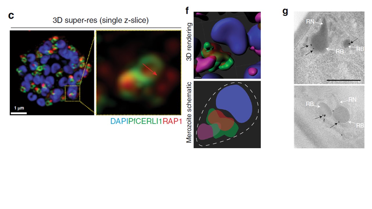
PfCERLI1 is peripherally-associated with the cytosolic face of the rhoptry membrane. c A single z-slice of the double bulbous rhoptries displaying the scheme that was used to measure the fluorescence intensity peaks (d) and diameter (e) of anti-HA and anti-RAP1 staining across the rhoptry (****p < 0.0001 by unpaired two-tailed t-test, n = 5 biological replicates, 1139 rhoptries measured for PfCERLI1HA and 1040 for RAP1, Error bars = SEM). f 3D rendered image of a free merozoite showing the nucleus (DAPI) localising at the basal surface, PfCERLI1 (HA) wrapping around RAP1 at the rhoptry bulb and RON4 localising in the rhoptry neck at the far apical tip. g Representative image of Compound 1 treated PfCERLI1HAGlmS schizonts that were fixed, labelled with anti-HA antibodies and probed with 18 nm colloidal gold secondary antibodies, before being imaged using transmission electron microscopy. White arrows mark rhoptry bulb (RB) and neck (RN), while black arrows mark PfCERLI1 foci. Scale bar = 500 nm.
Liffner B, Frölich S, Heinemann GK, Liu B, Ralph SA, Dixon MWA, Gilberger TW, Wilson DW. PfCERLI1 is a conserved rhoptry associated protein essential for Plasmodium falciparum merozoite invasion of erythrocytes. Nat Commun. 2020 11(1):1411.
Other associated proteins
| PFID | Formal Annotation |
|---|---|
| PF3D7_0210600 | cytosolically exposed rhoptry leaflet interacting protein, protein CERLI1 |