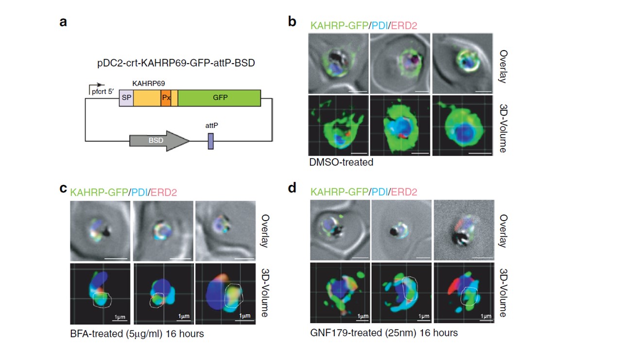
Confirmation of GNF179’s inhibition of protein export in P. falciparum blood stage parasites. a Vector used for assays shown in b–d. b–d Imaging of GFP reporter to the plasma membrane under the indicated compound treatments for 16 h. Parasites were fixed and stained with Hoechst 33342 (blue), α-GFP (green), α-ERD2 (red) and α-PDI (cyan) antibodies. Scale bars: 2 μm unless otherwise indicated. Overlay: DIC images merged with fluorescent channels. Quantification of colocalization between KAHRP-GFP and ERD2. we constructed a parasite strain that bears a fusion between the Knob-Associated Histidine Rich Protein and GFP56 (a). The chimeric gene, which consists of the first 69 amino acids of KAHRP containing the signal peptide (SP) and PEXEL motif (Px), was expressed from a pfcrt promoter and was integrated into the cg6 locus in Dd2-attB parasites using the attP × attB integrase system. In the absence of GNF179 the GFP reporter was trafficked to the PV as well as to the RBC cytosol, as shown by the GFP staining in and outside the PV surrounding the parasite (b). In contrast, treatment with brefeldin A resulted in accumulation of the GFP reporter in the parasite ER as evidenced when co-staining with an anti-PDI antibody (cyan) (c) or by using ER tracker (red). The white dotted outlines indicate overlap of green and cyan labels. We also observed some colocalization between the GFP and ERD2 signal (c) under brefeldin A treatment, though this correlation is lower than what is seen between GFP and PDI.
LaMonte GM, Rocamora F, Marapana DS, et al. Pan-active imidazolopiperazine antimalarials target the Plasmodium falciparum intracellular secretory pathway. Nat Commun. 2020;11(1):1780.
Other associated proteins
| PFID | Formal Annotation |
|---|---|
| PF3D7_0202000 | knob-associated histidine-rich protein |
| PF3D7_1353600 | ER lumen protein retaining receptor |