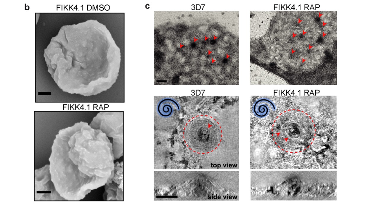
(b) SEM images of the surface of erythrocytes infected with DMSO or RAP-treated FIKK4.1::DiCre parasites. The experiment was repeated 2 times with similar results (scale bar - 1 μm). (c) Top panels: Electron micrographs of knobs on detergent treated, negatively-stained RBC ghosts from wild-type 3D7 and FIKK4.1KO schizonts imaged at low magnification. Red arrows indicate the position of knobs, which show up as circular dark patches on the membrane (scale bar - 200 nm). Bottom panels: Negative-stain electron tomography reveals typical structural features of the knob complex. In top views of knobs (XY), the knob coat is outlined with a red dashed line and the underlying knob spiral is indicated by red arrow heads. In these examples of knobs, the underlying spiral is left-handed (indicated by the blue spiral symbol) showing that the knob is pointing upwards from the plane of the grid. A side view (XZ) of the same knob shows that the height and diameter of the knobs in wild-type 3D7 schizonts FIKK 4.1 KO schizonts is similar. These knobs are pointing downwards and are compressed against the surface of the grid, hence more rings of the spiral structure are visible in the plane of the tomogram. Images are an average of 5 central slices of the tomogram (scale bar - 50 nm)
Davies H, Belda H, Broncel M, et al. An exported kinase family mediates species-specific erythrocyte remodelling and virulence in human malaria Nat Microbiol. 2020;10.1038/s41564-020-0702-4.