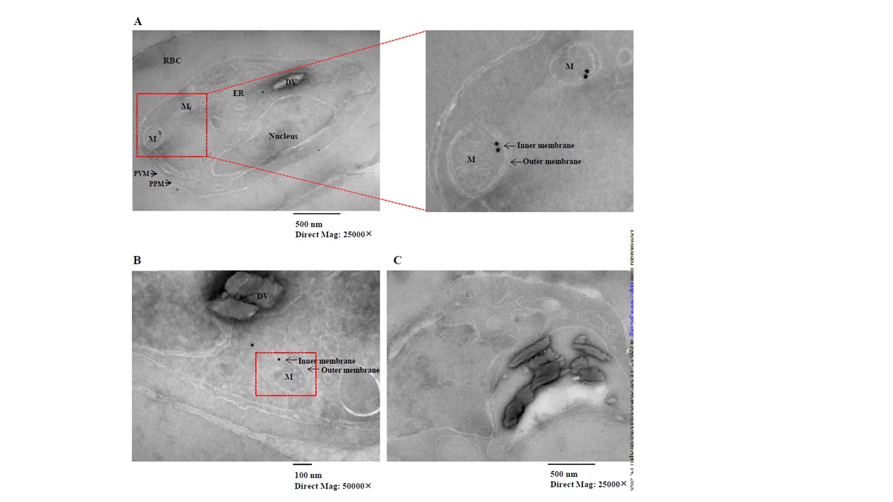
PfmtRPS18 is likely localized to the mitochondrial inner membrane. (A), (B) Images of D10-PfmtRPS18-3HA parasites. RBC, red blood cell. PVM, parasitophorous vacuole membrane. PPM, parasite plasma membrane. DV, digestive vacuole. ER, endoplasmic reticulum. M, mitochondrion. Black dots in red boxes indicate PfmtRPS18-3HA is localized to the mitochondrial inner membrane. (C) A control with omission of the primary antibody shows clear backgound. Mag, magnification. Although gold particles sometimes were present at other sites, the majority of them were found in the mitochondria at or very near the MIM (Figure 3A and 3B). In particular, quantification of gold particles from two biological experiments revealed the particles were localized to the mitochondria (at or near MIM) at 66.8%, the cytoplasm at 11.1%, the ER at 10%, the nucleus at 8.7% and others at 3.4%. Hence, our data indicate that PfmtRPS18 was likely associated with the MIM. As a negative control, omission of the primary antibody had a clean background (C). Together, these data suggest the Plasmodium mitoribosome is likely bound to the mitochondrial inner membrane, as in other organisms.
Ling L, Mulaka M, Munro J, Dass S, Mather MW, Riscoe MK, Llinás M, Zhou J, Ke H. Genetic ablation of the mitoribosome in the malaria parasite Plasmodium falciparum sensitizes it to antimalarials that target mitochondrial functions. J Biol Chem. 2020 Apr 9:jbc.RA120.012646.