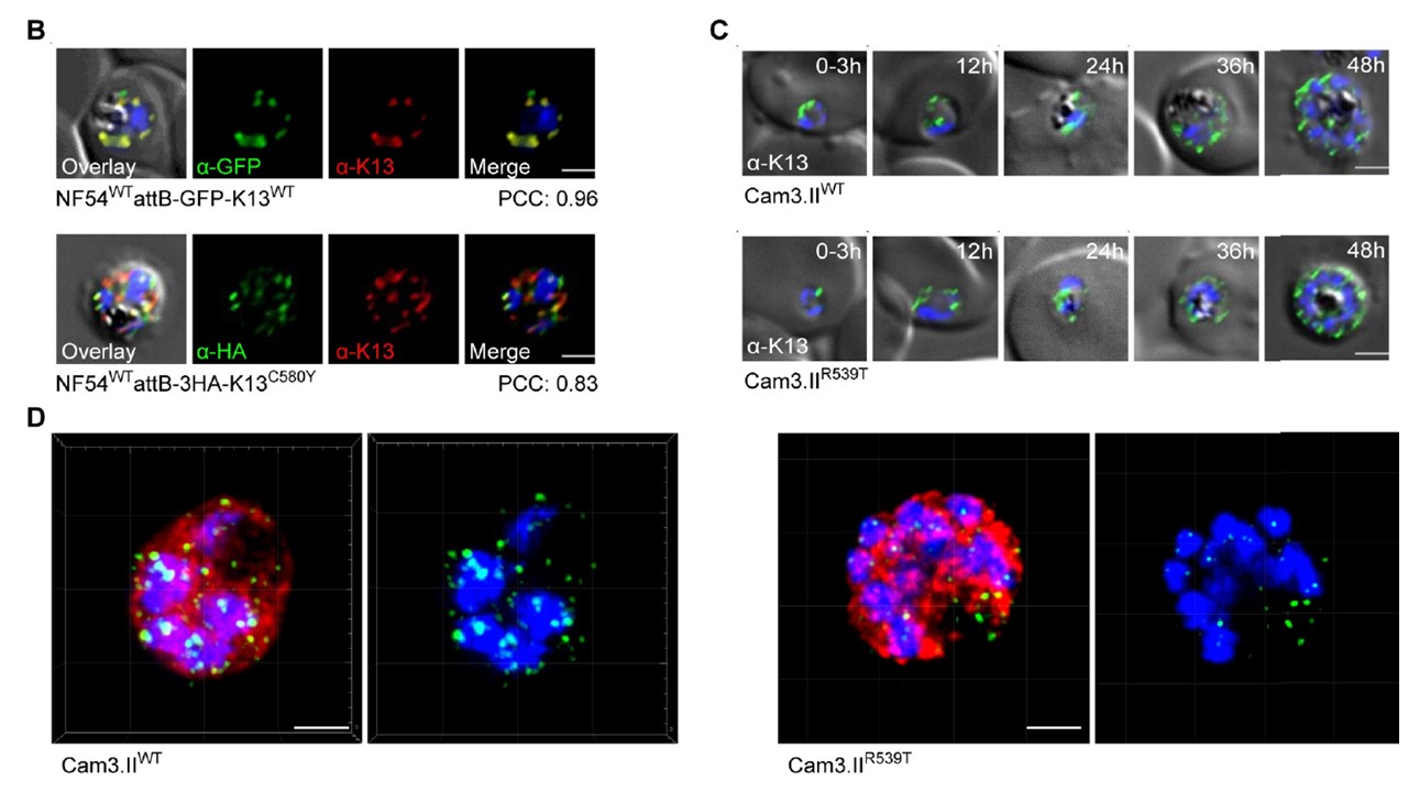
The P. falciparum artemisinin resistance determinant K13 localizes to the parasite ER and intracellular vesicles. (B) Immunofluorescence assay (IFA) images showing K13 localization in NF54WTattB-GFP-K13WT (top) and NF54WTattB-3HA-K13C580Y (bottom) trophozoites. Parasites were co-stained with the K13 E3 mAb and antibodies specific to GFP or HA. Pearson correlation coefficient (PCC) values indicate the degree of spatial co-localization between the two signals and were calculated by determining the fluorescence intensity correlations of Alexa Fluor 488 (anti-GFP or anti-HA) and 594 (K13 mAb). Nuclei were stained with DAPI (blue). Scale bars: 2 μm. (C) IFA images depicting K13 localization in Cam3.IIWT (top) and Cam3.IIR539T (bottom) parasites throughout asexual blood-stage development. Parasites were stained with the K13 E3 mAb. Sampling was performed every 12h, beginning with tightly synchronized 0–3 hpi ring-stage parasites. Scale bars: 2 μm.
Gnädig NF, Stokes BH, Edwards RL, Kalantarov GF, Heimsch KC, Kuderjavy M, Crane A, Lee MCS, Straimer J, Becker K, Trakht IN, Odom John AR, Mok S, Fidock DA. Insights into the intracellular localization, protein associations and artemisinin resistance properties of Plasmodium falciparum K13. PLoS Pathog. 2020 Apr 20;16(4):e1008482.